Figures & data
Figure 1. Transthoracic echocardiogram – subcostal window – showing a large circumferential pericardial effusion with right ventricular collapse
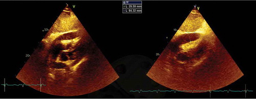
Figure 2. Positron emission tomography showing a focus of moderate hypermetabolism in the posterior region of the right pleura
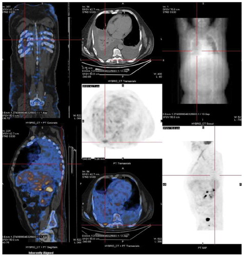
Figure 3. Pleural biopsy showing tuberculosis granuloma with a central caseous necrosis; magnification x100 and x200, respectively)
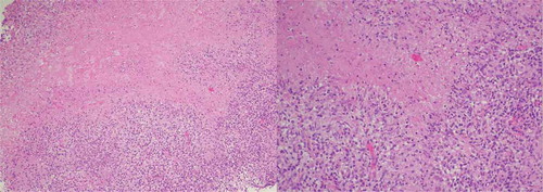
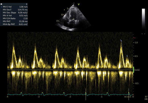
Figure 4. Tissue doppler imaging showing the relationship between lateral e’ and medial e’ velocities: Medial e’ > Lateral e’ suggestive of constrictive process (‘annulus reversus’)
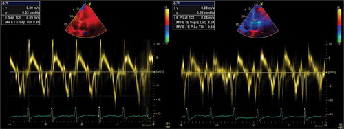
Figure 5. Pulsed wave Doppler showing an E/A ratio >2 suggestive of restrictive mitral inflow velocity