Figures & data
Figure 1. Magnetic resonance cholangiopancreatography
This is a coronal section of our patient’s Magnetic Resonance Cholangiopancreatography. This demonstrates an enlarged distal common bile duct. The duct actually enlarged by 3 mm compared to imaging 3 months prior, indicating a probable source of infection or cholangitis.
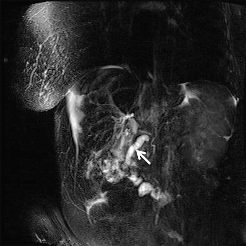
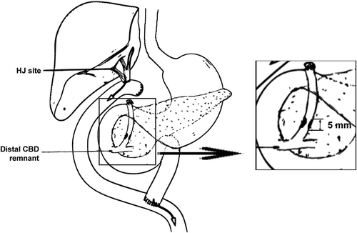
Figure 2. Patient anatomy with CBD stricture
This figure depicts our patient’s gastrointestinal tract following right lobe liver transplant connected via Roux-en-Y Hepaticojejunostomy. The enlarged rectangular portion highlights the remaining intrapancreatic common bile duct that is native to our patient. Note the 5 mm moderate stenosis in the distal portion, felt to be the source of our patient’s infection. CBD – Common Bile Duct; HJ Site – Anastomosis of Hepaticojejunostomy.