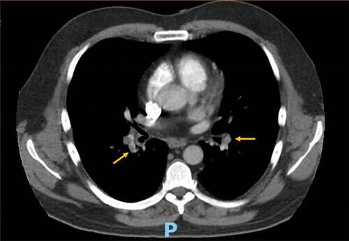
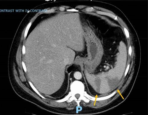
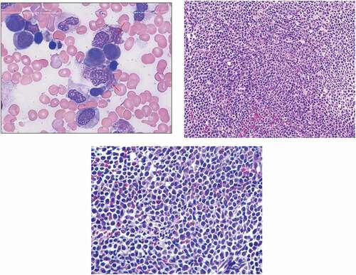
1,515
Views
0
CrossRef citations to date
0
Altmetric
Case Report

Acute promyelocytic leukemia presenting as recurrent venous and arterial thrombotic events: a case report and review of the literature

ORCID Icon, ORCID Icon, , &
Pages 832-838 | Received 01 Jun 2021, Accepted 24 Aug 2021, Published online: 15 Nov 2021

Figures & data

Table 1. Complete blood count with differential throughout clinical course

Figure 1. Computer tomography angiography of the chest demonstrating bilateral segmental lower lobe pulmonary embolism

Figure 1. Computer tomography angiography of the chest demonstrating bilateral segmental lower lobe pulmonary embolism

Table 2. Hypercoagulable workup

Figure 2. Computer tomography abdomen revealing wedge-shaped defects of the spleen consistent with infarcts

Figure 2. Computer tomography abdomen revealing wedge-shaped defects of the spleen consistent with infarcts

Figure 3. a.Bone marrow aspirate smear showing promyelocytes with ovoid to monocytoid nuclei, abundant cytoplasm with numerous pink, red or purple granules that obscure the nuclear outline. the cells contain numerous intertwining auer rods (arrows). b. The bone marrow biopsy showing hypercellularity with aggregates of promyelocytes (x200). c. High power image showing promyelocytes with relatively abundant cytoplasm and convoluted nuclei that are often eccentrically located (x400)

Figure 3. a.Bone marrow aspirate smear showing promyelocytes with ovoid to monocytoid nuclei, abundant cytoplasm with numerous pink, red or purple granules that obscure the nuclear outline. the cells contain numerous intertwining auer rods (arrows). b. The bone marrow biopsy showing hypercellularity with aggregates of promyelocytes (x200). c. High power image showing promyelocytes with relatively abundant cytoplasm and convoluted nuclei that are often eccentrically located (x400)

Table 3. Review of the literature of patients with acute promyelocytic leukemia (APL) presenting with thrombosis