Figures & data
Table 1. Cases of pancreatic carcinoma associated polymyositis and myopathy
Figure 1. MRI of patient’s lumbar spine. T2 image with solid arrows showing edema of paraspinal muscles. Outlined arrows indicated psoas muscles, which are non-edematous
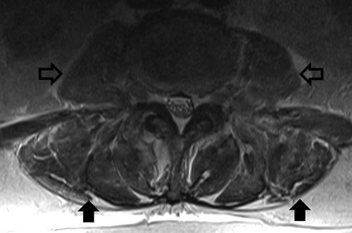
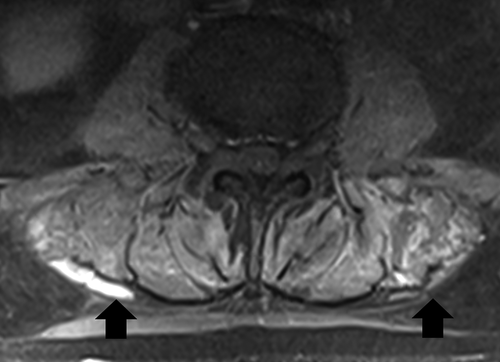
Figure 2. MRI of patient’s lumbar spine. T1 with contrast image with arrows indicating areas of enhancement in lumbar paraspinal muscles