Figures & data
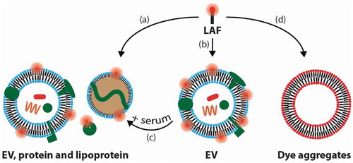
Figure 1. Schematic illustration of the proposed paths of the lipid-anchored fluorophore (LAF) used for labelling and tracking EVs that are discussed in this commentary.
(a) LAF labelling of non-EV serum components, that are often present in EV isolates, in addition to EV labelling. (b) LAF labelling of pure EVs. (c) Dissociation of LAFs from EVs into serum components. (d) Formation of LAF particles that exhibit low fluorescence due to self-quenching.