Figures & data
Text Box 1. Roundtable topics, moderators, and descriptions.
Table 1. Workshop pre-survey questions.
Table 2. Survey questions regarding EV biogenesis.
Table 3. Survey questions regarding EV uptake.
Table 4. Survey questions regarding current technologies for studying EVs.
Table 5. Summary of topics on which there is largely agreement, relative consensus, or clear lack of consensus; a set of specific recommendations are included.
Figure 1. EV Identification and size. Six questions regarding EV identification and sizing were administered in the post-workshop survey. For each question, participants’ answers are depicted horizontally on a Likert-scale from 0 to 10, with bubble size reflecting of the number of responders at each point on the scale. Most responders believe that there are multiple distinct pathways for vesicle biogenesis that result in heterogeneity in terms of size. Identifying vesicles from these pathways based on size, protein or lipid markers remains difficult.
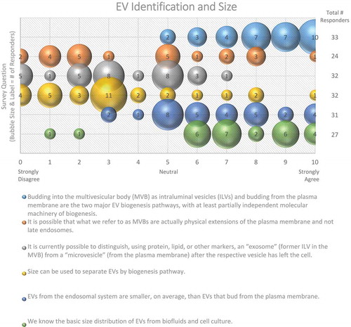
Figure 2. The average diameter of EVs (Excluding apoptotic bodies and other “macrovesicles”). In the post-workshop survey, participants were asked to choose from the three listed options. Responders believe that most EV populations are less than 150 nm in size. Those vesicles less than 100 nm in size are difficult to detect using techniques based on light scattering.
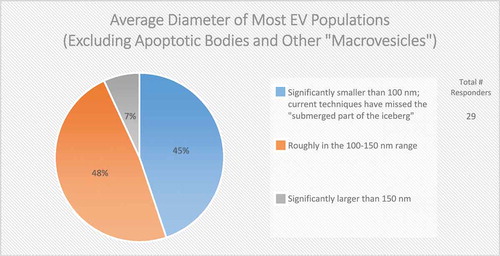
Figure 3. EV membrane topology. Two questions regarding EV membrane topology were administered in the post-workshop survey. For each question, participants’ answers are depicted horizontally on a Likert-scale from 0 to 10, with bubble size reflecting of the number of responders at each point on the scale. Responders are uncertain as to whether the lipid distribution of EV membranes is the same as the original cell membrane.
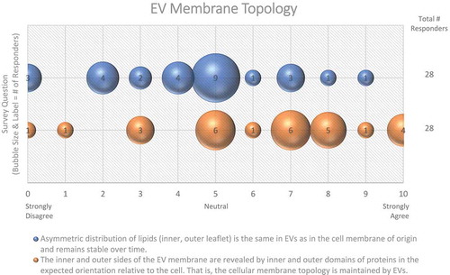
Figure 4. Membrane Involvement in EV cargo packaging. Two questions regarding the involvement of membranes in EV cargo packaging were administered in the Post-Workshop survey. For each question, participants’ answers are depicted horizontally on a Likert-scale from 0 to 10, with bubble size reflecting of the number of responders at each point on the scale. Responders are not sure whether miRNA or RNA cargo is specific to certain subtypes of EVs.
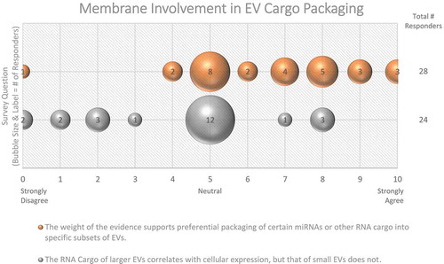
Figure 5. Importance of EVs for Life. Two questions regarding the importance of EVs for life were administered in the post-workshop survey. For each question, participants’ answers are depicted horizontally on a likert-scale from 0 to 10, with bubble size reflecting of the number of responders at each point on the scale. The majority of responders believe that EV production is necessary for cell and organism survival.
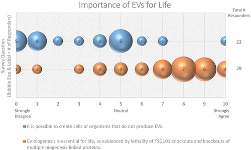
Figure 6. Requirements for EV Biogenesis. Three questions regarding requirements for EV biogenesis were administered in the post-workshop survey. For each question, participants’ answers are depicted horizontally on a likert-scale from 0 to 10, with bubble size reflecting of the number of responders at each point on the scale. Responders believe that the roles of lipid-raft domains, nSMase2, and the energetic requirements of EV biogenesis need to be further explored.
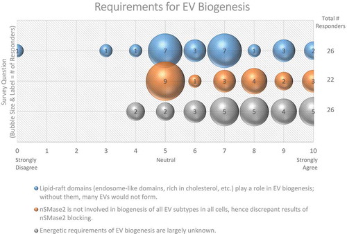
Figure 7. EV-cell fusion. Five questions regarding EV-cell fusion were administered in the post-workshop survey. For each question, participants’ answers are depicted horizontally on a Likert-scale from 0 to 10, with bubble size reflecting of the number of responders at each point on the scale. Responders agree that recipient cells internalize EVs from different cell types through endosomal uptake and acidification, and that proteins on the EV surface are responsible for fusion events. Survey participants are not sure how frequent EV-cell fusion events are in vivo.
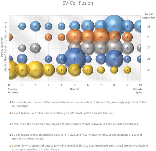
Figure 8. EV-cell interactions. In the post-workshop survey, participants were asked to rank order the most to least likely ways in which EVs interact with target cells. Answers are depicted in a heat map, with pink shades indicating a higher number of responders, and blue indicating a lower number of responders. Responders believe that EVs primarily interact with target cells by signalling through proteins displayed on the target-cell surface or endosomal lumen. Transferring functional RNA, proteins and lipids is seen as a secondary effect. Most believe that EVs are indirectly a form of nutrition or molecular recycling for recipient cells.
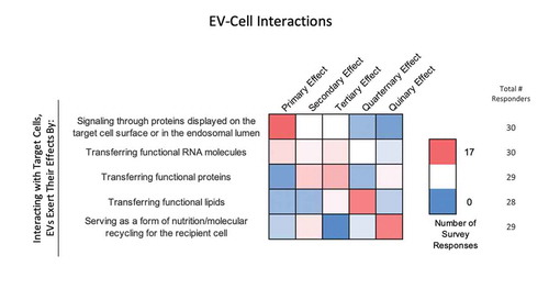
Figure 9. EV stability. Two questions regarding EV stability were administered in the post-workshop survey. For each question, participants’ answers are depicted horizontally on a Likert-scale from 0 to 10, with bubble size reflecting of the number of responders at each point on the scale. While EVs are physically stable, most survey participants believe that current technologies need to be improved to simultaneously measure the functional and physical stability of EVs.
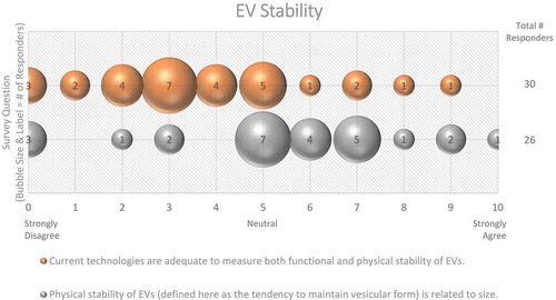
Figure 10. Storage of EVs. In the post-workshop survey, participants were asked to choose from five options whether or not they believe freeze-thawing causes damage to EVs. Responders agree that we do not know enough about how freeze-thawing affects EV stability, uptake, and functionality.
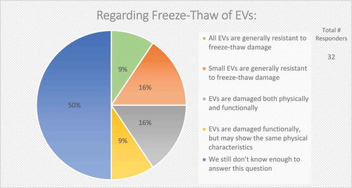
Figure 11. Bioactivity of EVs. Seven questions regarding the bioactivity of EVs were administered in the post-workshop survey. For each question, participants’ answers are depicted horizontally on a Likert-scale from 0 to 10, with bubble size reflecting of the number of responders at each point on the scale. Responders believe that the use of EVs for in vitro transfer experiments is time-dependent, that dose–response studies are important, and that EVs have a greater functional impact in the local tissue environment. Survey participants are undecided on how to determine and identify bioactive EVs. The survey reveals the need for improved technology for the study of EV-cell fusion.
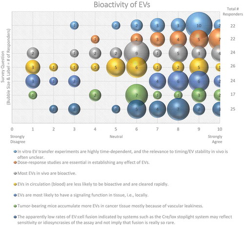
Figure 12. Current particle tracking technologies. Three questions regarding current particle tracking technologies were administered in the post-workshop survey. For each question, participants’ answers are depicted horizontally on a Likert-scale from 0 to 10, with bubble size reflecting of the number of responders at each point on the scale. Survey participants require improved, non-biased technologies for determining EV size. The use of lipid dye can cause experimental artefacts.
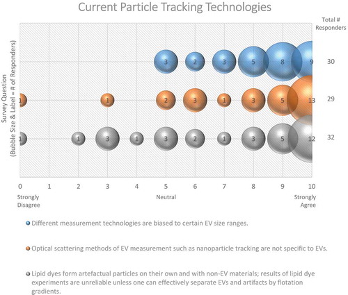
Figure 13. Current opinion on EV-flow cytometry. In the post-workshop survey, participants were asked to choose between two options regarding the current status of applying flow cytometry to the study of EVs. Almost all responders to this question call for specialized equipment, reagents and expertise to characterize single EVs through flow cytometry.
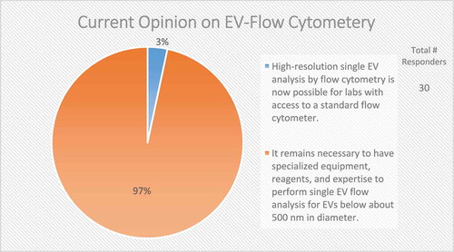
Figure 14. Fluorescent labelling of EVs. Three questions regarding fluorescent labelling of EVs were administered in the post-workshop survey. For each question, participants’ answers are depicted horizontally on a Likert-scale from 0 to 10, with bubble size reflecting of the number of responders at each point on the scale. Survey question participants acknowledge that better dyes and reagents are needed to study EVs using flow cytometry. Still, using fluorescent flow cytometry to study EVs provides better resolution than scatter.
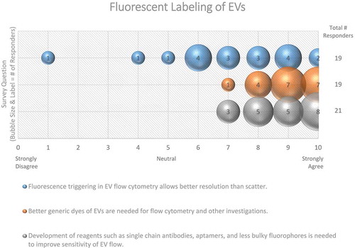
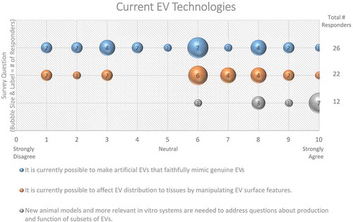
Figure 15. Current EV Technologies. Three questions regarding current EV technologies were administered in the post-workshop survey. For each question, participants’ answers are depicted horizontally on a Likert-scale from 0 to 10, with bubble size reflecting of the number of responders at each point on the scale. Responders agree that new animal and in vitro models are needed to address questions concerning EV production and function. Survey participants are not sure whether artificial EVs can mimic genuine EVs, or that manipulation of EV surface features will affect biodistribution.