
Figures & data
Table 1. Patient characteristics at time of inclusion
Table 2. HRCT findings at time of inclusion
Table 3. Histological diagnosis based on cryobiopsies
Table 4. Clinical diagnosis after a multidisciplinary team discussion
Figure 1. 71-year-old male referred for dyspnea. HRCT showing reticulation, ground glass opacity and traction bronchiectasies with basal predominance. Cryobiopsies showing patchy fibrosis, fibroblastic foci and chronic inflammation. The patient was diagnosed with idiopathic pulmonary fibrosis, high confidence
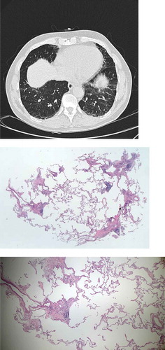
Figure 2. 51-year-old male referred for increasing dyspnea and cough. HRCT showing diffuse reticulation, ground glass opacity, traction bronchiectasies and consolidations. Cryobiopsies showing chronic inflammation, fibrosis and granulomas/giant cells. The patient was diagnosed with chronic hypersensitivity pneumonitis
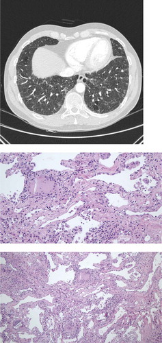
Table 5. Cryobiopsy complications