Figures & data
Figure 1. (a) Chest radiograph: multiple bilateral mid and lower zone airspace opacities. (b) Chest radiograph: pneumatocele with air fluid level in right lower lobe. (c) Follow up chest radiograph: resolution of pneumatocele in the follow up CXR.
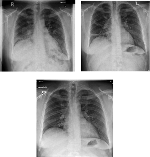
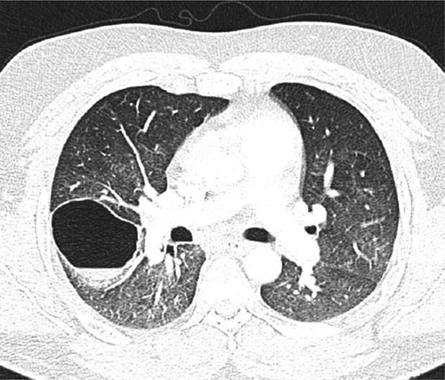
Figure 2. CT Chest: Right lower lobe pneumatocele with air fluid level. Other findings included bilateral ground glass changes.