Figures & data
Figure 1. Chest CT scan, evidence of S3 consolidation, and cavitation of the left hemithorax.
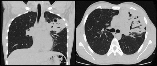
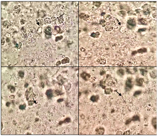
Figure 2. Lophomonas (black arrow) detected in the bronchoalveolar lavage fluid by microscopic observation: direct smear (×100).
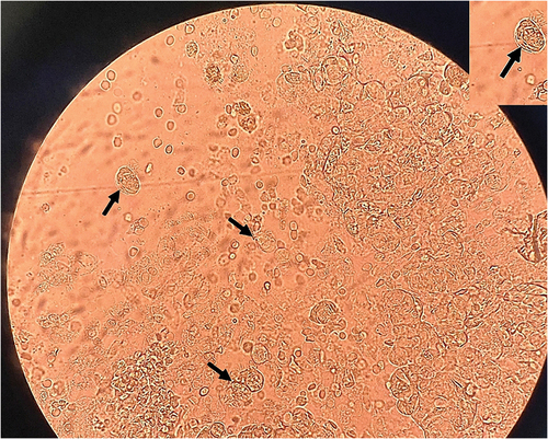
Figure 3. Chest CT scan, evidence of a cavity with content compatible with mycetoma, and multiple bronchiectasis in S1 and 2.
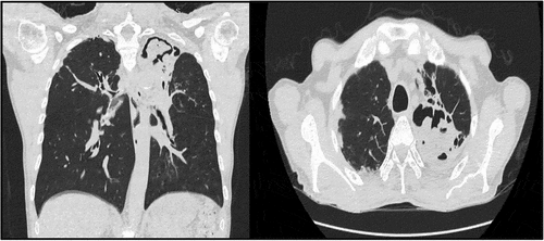
Figure 4. Sequential microscopic examination of Lophomonas (black arrow) in the direct smear of bronchial aspirate.