Figures & data
Figure 1. Image of the EUS echo-endoscope (a) and the EBUS echo-endoscope (b).
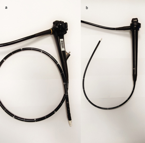
Figure 2. Overview of structures that can be reached by EBUS, EUS/EUS-B or both.
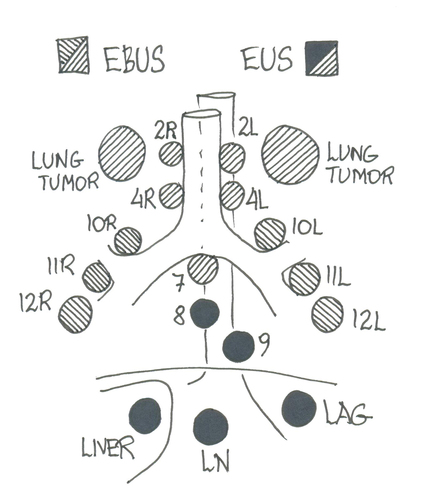
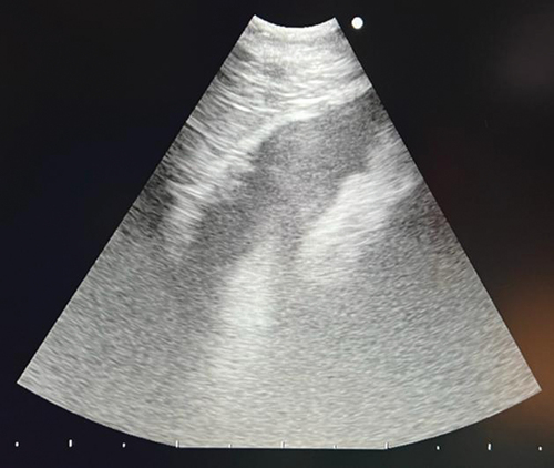
Figure 3. EUS-B image of mediastinal lymph node station 7.
Figure 4. EUS-B image of mediastinal lymph node station 4 R.
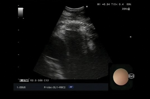
Figure 5. EUS-B image of mediastinal lymph node station 4 L.
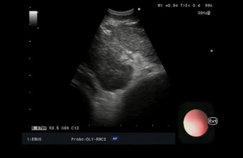
Figure 6. EUS-B image of mediastinal lymph node station 8.
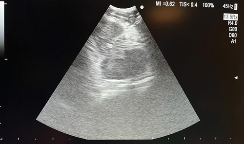
Figure 7. EUS-B image of a lung tumor in the left upper lobe being biopsied.
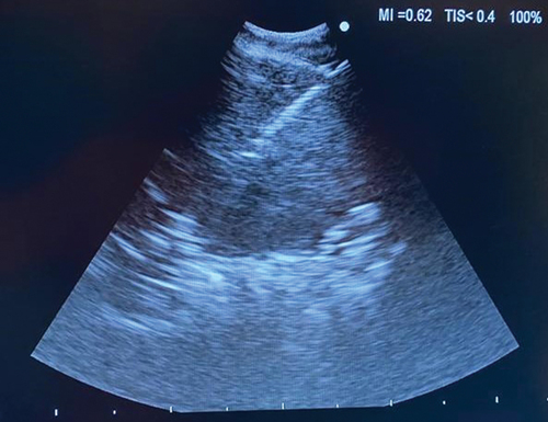
Figure 8. EUS-B image of the liver.
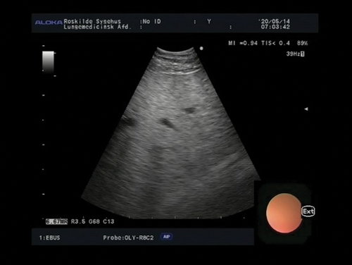
Figure 9. EUS-B image of the left adrenal gland.
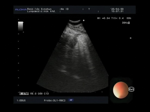
Figure 10. EUS-B image of a retroperitoneal lymph node being biopsied.
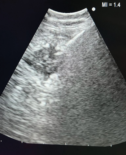
Figure 11. EUS-B image of a pericardial effusion.