Figures & data
Figure 1. Chest X-ray showing bilateral scattered interstitial consolidations.
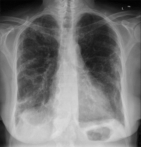
Figure 2. High-resolution computed tomography revealed interstitial changes with irregular reticulation (left) and basal traction bronchiectasis and subpleural consolidation (right).
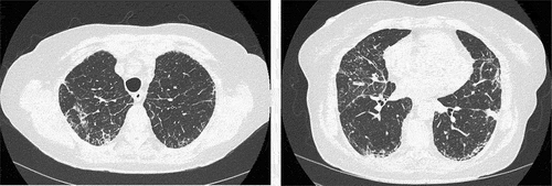
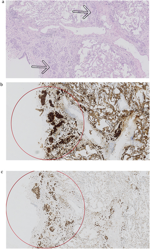
Figure 3. Histopathological evaluation of lung cryobiopsy. (a) Interstitial, intralymphatic solid nests of epithelial tumour cells (arrows) (×50), (b) positive for CK7 (×50), and (c) positive for GATA3 (×50).