Figures & data
Table 1. Organisms and pathogens used by Infravec2 consortium members who contributed to these guidelines. This guidance document is informed by the experience of the contributors and as such particularly reflects requirements for work with insects (mainly mosquitoes), which can be endemic, imported or exotic species. Moreover, these species can be genetically modified (e.g. lines containing a docking site, mutant for a gene, overexpressing anti-pathogen effectors, bearing gene drive systems, etc.) and/or be transinfected with endosymbionts, e.g. Wolbachia.
Table 2. Summary of hazard groups (HG) and containment levels (CL) for the purpose of this document.
Figure 1. Layout for a CL2 insectary. The main room is either humidity and temperature controlled (in this case, electric equipment must be placed outside the humidity room, in the antechamber or in a room close by) or equipped with environmental chambers. Air showers/curtains can be useful upon exit to avoid escapes. Benching and shelving can be stacked up to gain storage space.
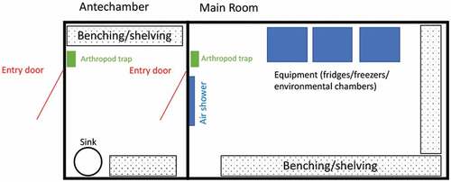
Figure 2. Stacked shelves housing breeding pans and cages in a humidity- and temperature-controlled room.
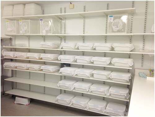
Figure 3. Examples of environmental/climatic chamber with humidity, temperature, and light control. (a, b) Climatic chamber connected to a water container for water supply and to a waste container to collect wastewater. (c) Walk in climatic chamber.

Figure 4. Room with stereomicroscopes and material for mosquito manipulation, such as dissection and injection.
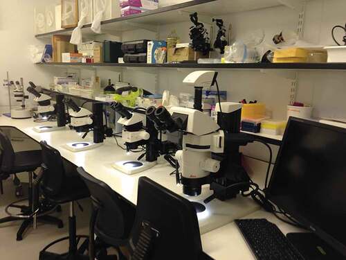
Figure 5. Primary containers for non-infected mosquitoes. Left panel, breeding pan covered with a weighted net for mosquito larvae. Adults can emerge in pans and are further aspirated. Right panel, typical cage for adult mosquitoes with plastic and meshed panels plus a mesh sleeve allowing access.
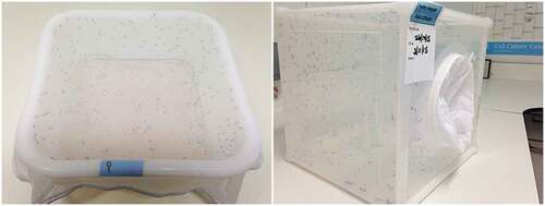
Figure 6. Example of CL2 insectaries dedicated to mosquito infection with HG2 arboviruses. The room is equipped with a class II MSC, to prepare the infectious blood and blood capsules, a glove box, which is a sealed container allowing user to manipulate flying arthropods inside the box without breaking containment, a bench with stereomicroscopes for mosquito dissection, environmental chambers, a freezer to freeze all solid waste before taking out for autoclaving, a sink to dispose of chemically inactivated waste, a UV light trap, a fridge and storage shelves/furnitures.
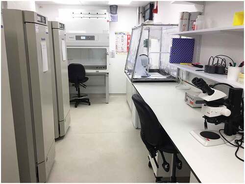
Figure 7. Example of primary and secondary containers. (a) Small and secured primary container with a netted lid. (b) Primary containers in secondary container for transport. (c) Primary containers in meshed cage inside environmental chamber.
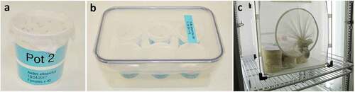
Figure 8. Mosquito blood feeding with non-infectious or infectious blood meals. (a) The blood is poured into capsules. When blood is to be spiked with pathogen, this step should be performed in an MSC. (b) Feeding capsule filled with blood and covered by an artificial membrane. (c) Blood capsule connected to the heating unit. (d, e) non-infectious blood feeding of mosquitoes in cage. (f) Infectious blood feeding of mosquitoes in small and secured primary containers.
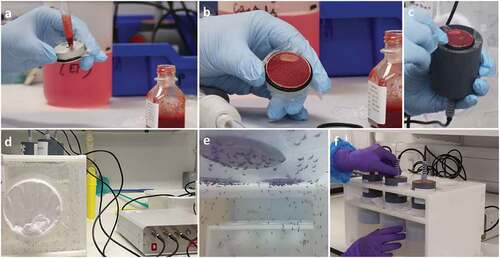
Figure 9. Enclosed feeding device system for Culicoides. (a) Primary container ‘netted feeding chamber’ where Culicoides will be kept during blood meal. (b) Secondary container, ‘infectious blood reservoir chamber’ in a water bath and magnetic stirrer with temperature probe. (c) Transport box for movement of infectious items between rooms.
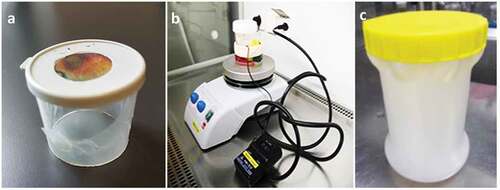
Figure 10. Layout of a CL3 insectary. Access to the CL3 area is via a lobby divided (by a door or simply by a mark on the floor) into a clean (entrance side) and a dirty (CL3 corridor side) area. After the lobby, a corridor can give access to insectary room(s) and a cell culture room which can be used to prepare and store pathogens. Mechanisms to avoid simultaneous opening of doors should be put in place. A double-ended autoclave allows autoclaving waste from the CL3 area and removal of autoclaved waste from outside the CL3 area. Store and equipment rooms are optional. Chemically inactivated liquid waste can be autoclaved or discarded using an effluent treatment system.
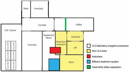
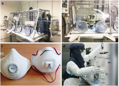
Figure 11. Protective equipment to work with HG3 pathogens. (a) HEPA-filtered isolator working under negative pressure for manipulation of HG3 infectious material which can be aerosol transmitted. A hatch allows material entry while work is ongoing. Height is adjustable. (b) Incorporated stereomicroscope to dissect infected mosquitoes in HEPA-filtered isolator. (c, d) Filtering face piece (FFP) 3 masks or Purifying Air Powered Respirators (PAPR) that can be used in CL3 in place of a HEPA-filtered isolator.