Figures & data
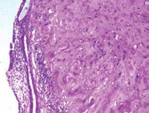
Figure 1: Medium power magnification view of the histology of the tumor in case 1, showing cords of epithelioid cells in a hyalinised background invading deep into the myometrium. Some of the epithelioid cells display clear cytoplasm. Mitotic activity can also be seen. Haematoxylin and eosin, 100 × magnification.
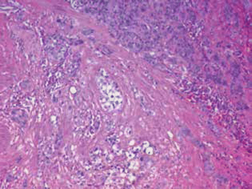
Figure 2: Medium power magnification view of the histology of the tumor in case 2, showing a nodule of mostly hyalinised tumor. Haematoxylin and eosin, 100 × magnification.