Figures & data
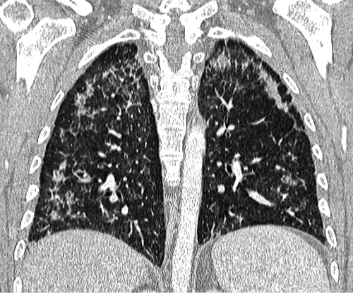
Figure 1: Biochemical marker response. AFP = alpha-fetoprotein; HCG = human chorionic gonadotropin; LDH = lactate dehydrogenase.
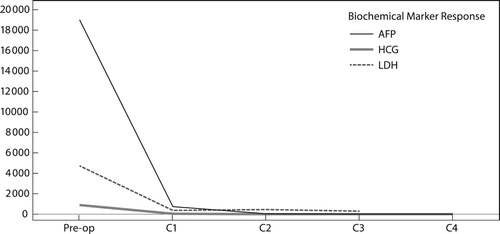
Figure 2: (a) Initial and (b) follow-up CT scan: upper lungs axial CT slices (lung windows): (a) There is intra- and inter-lobular septal thickening with associated ground-glass opacification and an incidental small-volume pneumomediastinum, the aetiology of which is not clear, possibly related to an air leak from the adjacent lung pathology. The lung changes are consistent with an NSIP pattern of lung injury or an organising pneumonia. (b) Interval progression is noted in terms of severity as well as area of the lung involved. There is now diffuse ground-glass opacification of the lungs as well as established fibrosis with regions of honeycombing. The large ground-glass component suggests an active cellular component to the disease and the radiological picture favours a mixed cellular fibrotic NSIP pattern of lung injury. The pneumomediastinum progressed and there is a left-sided pneumothorax.
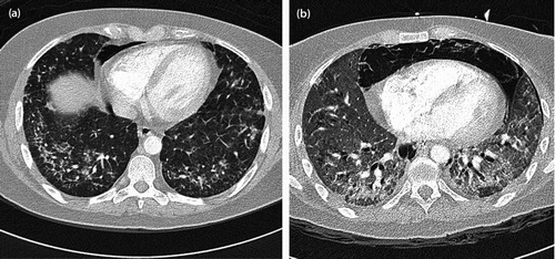
Figure 3: Coronal CT image (lung window): the image demonstrates the predominant peripheral distribution of changes with relative sub-pleural sparing as well as the lack of a clear apico-basal gradient.