Figures & data
Figure 1. Position of the patient and location of the perforator
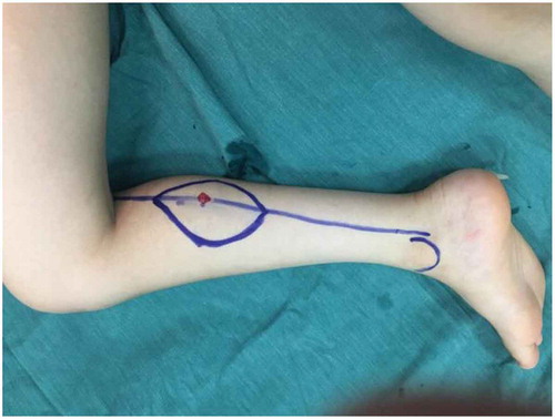
Figure 2. Medial incision and identification of the perforator
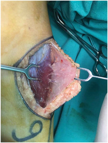
Figure 3. Dissection of the pedicle
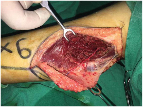
Figure 4. Direct closure of the donor site
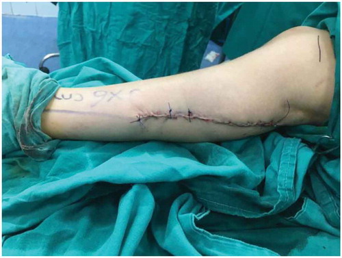
Figure 5. Skin graft of the donor site
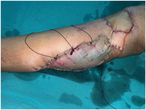
Figure 6. Case 1. a: Palmer surface of right hand showing soft tissue defect. b: Dorsal surface of right hand showing soft tissue defect. c: Flap marking. d: Anastomosis site. e: Dissection of the perforator. f: Dissection of the pedicel. g: Flap in-setting Palmer surface. h: Flap in-setting dorsal surface. i: Donor side closure. j: Patient after 1 week. k: Patient after 1 month. l: Patient after 3 months. m: Patient after separation of index finger. n: Patient during separation of middle and ring fingers showing the perforator. o: Patient after 6 months. p: Patient after 6 months. q: Patient after 6 months. r: Patient after 8 months
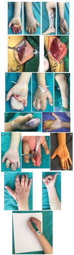
Table 1. Summary of the data of the patients included in this study
Figure 7. Case 2. a: Pre-operative photo showing the defect. b: Intraoperative photo showing the defect. c: Flap marking. d: Perforator dissection. e: Flap elevation. f: Flap insetting. g: Patient after 2 weeks. h: Patient after 2 months with good color match and thickness. i: Patient after 3 months. j: Donor side hypertrophic scar
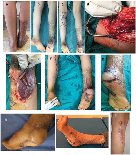
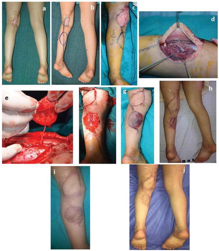
Figure 8. Case 3. a: The defect in the left knee. b: Marking of the flap. c: Excision of the defect. d: Dissection of the perforator. e: Flap elevation. f: Flap insetting. g: Patient after 1 week. h: Patient after 1 month. i: Patient after 5 months. j: Patient after 11 months with good color match and thickness