
Figures & data
Figure 1. Surgical steps of case # 6 (right eye with TRD and ERM of 49-year-old female patient suffering type 1 diabetes for 14 years) in (group A) (A) Injection of the vital dye under the PFCL. (B) Bimanual dissection of fibrovascular membranes. (C) after removal of fibrovascular proliferations.
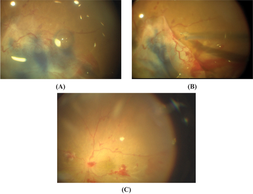
Figure 2. surgical steps of case # 5 (left eye with TRD and ERM of 51-year-old male patient suffering type 1 diabetes for14 years) in bimanual PPV group (group B): (A) Injection of the vital dye over the posterior pole. (B) Stained fibrovascular proliferations. (C) Bimanual dissection of the preretinal proliferation with intraoperative bleeding.
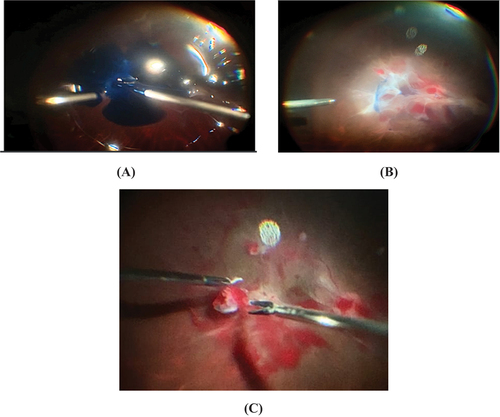
Table 1. Comparison between the two studied groups according to time of membrane removal and time of surgery.
Table 2. Comparison between the two studied groups according to intraoperative bleeding.
Figure 3. Comparison between the two studied groups according to iatrogenic breaks.
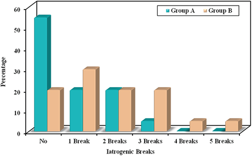
Table 3. Comparison between the two studied groups according to post-operative complication.
Table 4. Comparison between the different studied periods according to BCVA in each group.