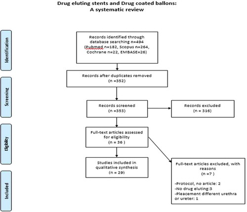
Figures & data
Table 1. Eligibility criteria of the systematic review
Table 2. Comparison of the experimental (in vitro) trials of DES/DCS
Table 3. Comparison of the clinical (in vivo) trials of DES/DCS
Table 4. Comparison of the clinical trials (in vivo) of DCBs
Antimisiaris SG, Siablis D, Liatsikos E, et al. Liposome-Coated Metal Stents: an in Vitro Evaluation of Controlled-Release Modality in the Ureter. J Endourol. 2000;14(9):743–747. Cadieux PA, Chew BH, Knudsen BE, et al. Triclosan loaded ureteral stents decrease proteus mirabilis 296 infection in a rabbit urinary tract infection model. J Urol. 2006;175(6):2331–2335. Barros AA, Oliveira C, Reis RL, et al. Ketoprofen-eluting biodegradable ureteral stents by CO2 impregnation: in vitro study. Int J Pharm. 2015;495(2):651–659. Barros AA, Oliveira C, Reis RL, et al. In Vitro and Ex Vivo Permeability Studies of Paclitaxel and Doxorubicin From Drug-Eluting Biodegradable Ureteral Stents. J Pharm Sci. 2017;106(6):1466–1474. Chew BH, Cadieux PA, Reid G, et al. Second Prize : in-Vitro Activity of Triclosan-Eluting Ureteral Stents against Common Bacterial Uropathogens. J Endourol. 2006;20(11):949–58. Elayarajah E, Rajendran R, Venkatrajah B, et al. Biopolymer tocopherol acetate as a drug carrier to prevent bacterial biofilm formation on silicone ureteral stents. Int J Pharm Sci Rev Res. 2011;7:96–103. Johnson JR, Johnston BD, Kuskowski MA, et al. In vitro activity of available antimicrobial coated foley catheters against escherichia coli, including strains resistant to extended spectrum cephalosporins. J Urol. 2010;184(6):2572–2577. Kotsar A, Isotalo T, Uurto I, et al. Urethral in situ biocompatibility of new drug-eluting biodegradable stents: an experimental study in the rabbit. BJU International. 2009;103(8):1132–1135. Ma X, Xiao Y, Xu H, et al. Preparation, degradation and in vitro release of ciprofloxacin-eluting ureteral stents for potential antibacterial application. Mater Sci Eng C Mater Biol Appl. 2016;66:92–99. El-Nahas AR, Lachine M, Elsawy E, et al. A randomized controlled trial comparing antimicrobial (silver sulfadiazine)-coated ureteral stents with non-coated stents. Scand J Urol. 2018;52(1):76–80. Cadieux PA, Chew BH, Nott L, et al. Use of triclosan-eluting ureteral stents in patients with long-term stents. J Endourol. 2009;23(7):1187–1194. Cauda F, Cauda V, Fiori C, et al. Heparin Coating on Ureteral Double J Stents Prevents Encrustations: an in Vivo Case Study. J Endourol. 2008;22(3):465–472. Chew BH, Davoudi H, Li J, et al. An In Vivo Porcine Evaluation of the Safety, Bioavailability, and Tissue Penetration of a Ketorolac Drug-Eluting Ureteral Stent Designed to Improve Comfort. J Endourol. 2010;24(6):1023–1029. Cirioni O, Ghiselli R, Minardi D, et al. RNAIII-inhibiting peptide affects biofilm formation in a rat model of staphylococcal ureteral stent infection. Antimicrob Agents Chemother. 2007;51(12):4518–4520. Han K, Park J-H, Yang S-G, et al. EW-7197 eluting nano-fiber covered self-expandable metallic stent to prevent granulation tissue formation in a canine urethral model. PLoS One. 2018;13(2):e0192430. Kallidonis P, Kitrou P, Karnabatidis D, et al. Evaluation of zotarolimus-eluting metal stent in animal ureters. J Endourol. 2011;25(10):1661–16617. Kim KY, Park J-H, Kim DH, et al. Sirolimus-eluting Biodegradable Poly- l -Lactic Acid Stent to Suppress Granulation Tissue Formation in the Rat Urethra. Radiology. 2018;286(1):140–148. Kotsar A, Nieminen R, Isotalo T, et al. Preclinical evaluation of new indomethacin-eluting biodegradable urethral stent. J Endourol. 2012;26(4):387–392. Kram W, Rebl H, Wyrwa R, et al. Paclitaxel-coated stents to prevent hyperplastic proliferation of ureteral tissue: from in vitro to in vivo. Urolithiasis. 2020;48(1):47–56. Krambeck AE, Walsh RS, Denstedt JD, et al. A novel drug eluting ureteral stent: a prospective, randomized, multicenter clinical trial to evaluate the safety and effectiveness of a ketorolac loaded ureteral stent. J Urol. 2010;183(3):1037–1042. Krane LS, Gorbachinsky I, Sirintrapun J, et al. Halofuginone-coated urethral catheters prevent periurethral spongiofibrosis in a rat model of urethral injury. J Endourol. 2011;25(1):107–112. Liatsikos EN, Karnabatidis D, Kagadis GC, et al. Application of paclitaxel-eluting metal mesh stents within the pig ureter: an experimental study. Eur Urol. 2007;51(1):217–223. Lin Y-C, Liu K-S, Lee D, et al. In Vivo and In Vitro Elution of Analgesics from Multilayered Poly(D,L)-lactide- co -glycolide Nanofibers Incorporated Ureteral Stents. J Nanomater. 2018;2018:4943210. Mendez-Probst CE, Goneau LW, MacDonald KW, et al. The use of triclosan eluting stents effectively reduces ureteral stent symptoms: a prospective randomized trial. BJU International. 2012;110(5):749–754. Shin JH, Song H-Y, Choi CG, et al. Tissue Hyperplasia: influence of a Paclitaxel-eluting Covered Stent—Preliminary Study in a Canine Urethral Model. Radiology. 2005;234(2):438–444. Wang Z-X, Hong B-F, Zhang X, et al. New biodegradable drug-eluting stents for urethral strictures in a rabbit model. J Bioact Compat Pol. 2011;26(1):89–98. Barbalias D, Lappas G, Ravazoula P, et al. Evaluation of the distribution of paclitaxel after application of a paclitaxel-coated balloon in the rabbit urethra. Journal of Endourology. 2018;32(5):381–386. Liourdi D, Kallidonis P, Kyriazis I, et al. Evaluation of the distribution of paclitaxel by immunohistochemistry and nuclear magnetic resonance spectroscopy after the application of a drug-eluting balloon in the porcine ureter. J Endourol. 2015;29(5):580–589.