Figures & data
Figure 1. Phylogenetic tree based on Neighbour Joining method analysis of 533 bp sequence of the cytochrome c oxidase subunit I (COI) gene. The green spots and the number at each node indicate the bootstrap support. * indicates the sequence from this case (GenBank MH069729).
Figure 2. Leptometopa latipes puparium in ventral (A), dorsal (B) and lateral (C) view (scale bar 500 µm). Puparium details: Posterior anal region (D), anal plate (E), intersegmental spicules (F), posterior spiracle (G, H) and anterior spiracle (I) (scale bar 100 µm).
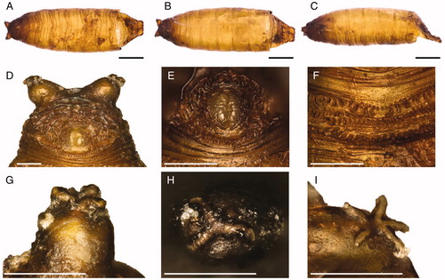
Figure 3. Leptometopa latipes puparium details: anal plate (A), intersegmental spicules (B), posterior spiracle (C, D) and filaments emanating from perispiracular glands (E), anterior spiracle (F).
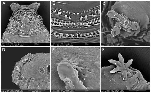
Figure 4. Leptometopa latipes adults head details. Female oral region (A), male bristles (B) and male antennae (C) (scale bar 100 µm).
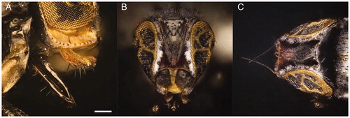
Figure 5. Leptometopa latipes adults details: thorax (A) and wing (B) (scale bar 100 µm).
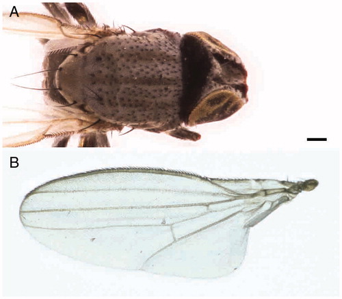
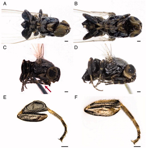
Figure 6. Leptometopa latipes adults. Male in ventral (A) and lateral (C) view. The arrow shows the yellowish ring in the foreleg. Female in ventral (B) and lateral (D) view. L. latipes hind tibia: male (E) and female (F) (scale bar 100 µm).