Figures & data
Figure 1. Laser scanning of the tooth specimen (A) and the 3D model of the scanned specimen (B).
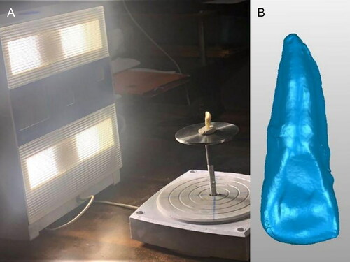
Figure 2. Fragmented tooth samples were obtained using a mortar and pestle.
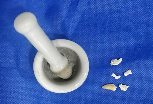
Figure 3. (A) Scanning of the fragmented tooth pieces using optical scanner. (B) 3D model obtained in Standard Tessellation Language (STL) format. (C) Multi-point registration performed manually, fragmented.
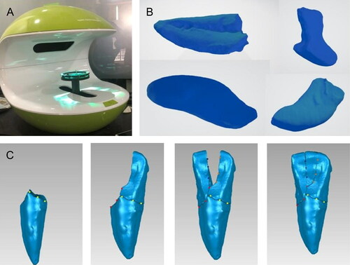
Figure 4. Models of reconstructed teeth which were printed using tooth printed by stereolithography technique (SLA) and tooth printed by fused deposition modelling technology (FDM) techniques (left to right, respectively).
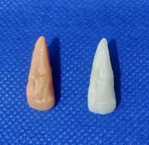
Table 1. Linear odontometric measurements of the reference teeth and 3D printed replicas to evaluate.
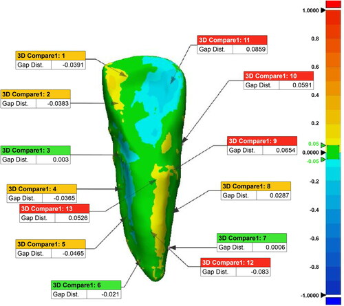
Figure 5. Qualitative congruency analysis performed on images of a reference tooth and a reconstructed tooth specimen.