Figures & data
Figure 1. Postmortem (PM) dental elements identifiers on examined bodies (22 PM odontograms).
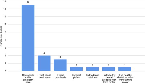
Figure 2. Postmortem dental elements identifiers (%) detected on the 2 D head X-rays performed in all the victims.
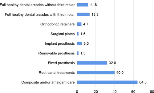
Figure 3. Antemortem files components.
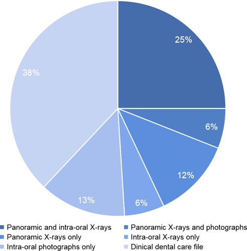
Figure 4. First-line postmortem 2 D radiographs, no dental care, complete arches few (mostly morphometric) data. On the right, there are only two orthodontic retainers (arrows).
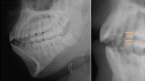
Figure 5. Antemortem (top) and postmortem (bottom) with orthognathic surgery osteosynthesis material.
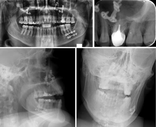
Figure 6. Antemortem radiological record (top) and 2 D postmortem face and profile images (bottom) with several strong markers: implant (arrow), anterior bridge (arrowhead), apical cementosis (star).
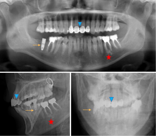
Figure 7. Antemortem X-ray record by bite-wings images, numerous amalgam and composite fillings (left and middle), postmortem image of first intention (right) superimposition of coronary radiopaque images, in this case intra-oral images are indispensable.
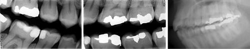
Figure 8. Panoramic reconstructed in thick section from a CT scan “head bone” volume.
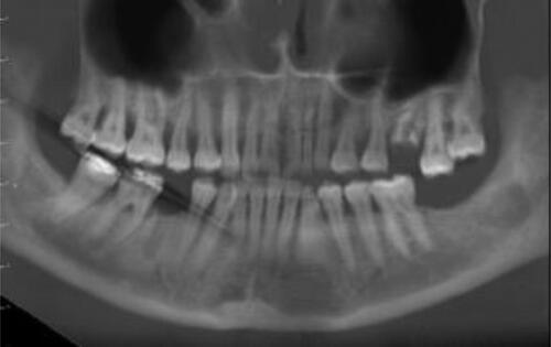
Figure 9. Panoramic view in maximum intensity projection mode.
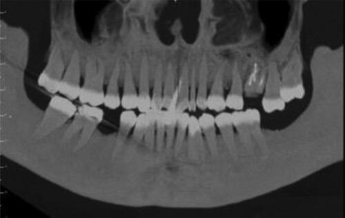
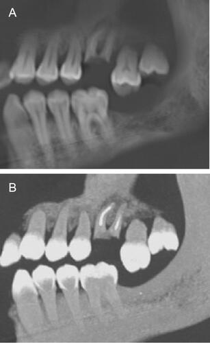
Figure 10. (A) Left side, first maxillary molar in the form of roots with endodontic treatment-egression second molar. (B) Maximum intensity projection mode in idem.