Figures & data
Figure 1. Plastic bags in which the body parts were found at the crime scene.
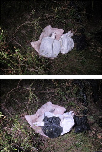
Figure 2. Decomposed body parts received at the Medico-Legal Institute.
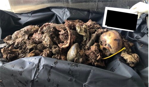
Figure 3. Illustration of the skeleton showing the locations of the injuries.
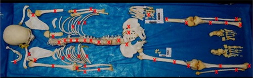
Figure 4. Sharp force and blunt force trauma on the maxilla, mandible, and clavicle.
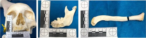
Figure 5. Sternum showing complete section with loss of the lower third of the manubrium. A rare anatomical variation was also seen at this site, a so-called suprasternal or episternal bone (see the red circle).
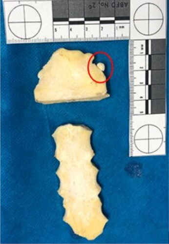
Figure 6. Several false starts with eversion of the bone margins on both scapulas and the proximal epiphysis of the left ulna (see the red circles).
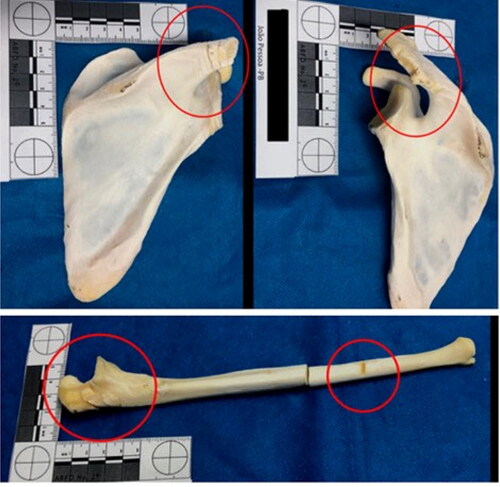
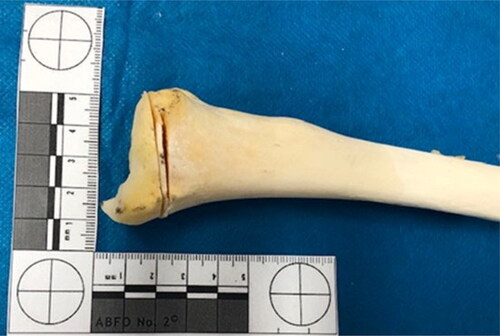
Figure 7. Sharp and blunt force trauma on the humerus and radius.
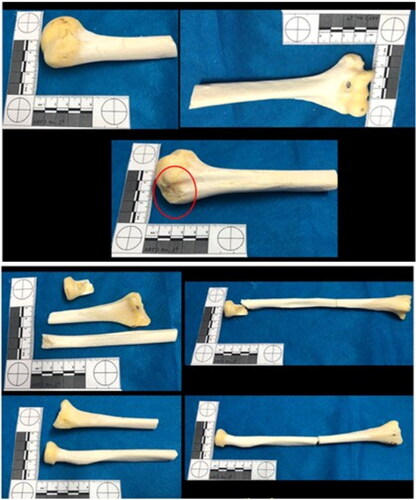
Figure 8. The spine mainly showed sharp force trauma on all its segments and cut impact marks (see the red circles).
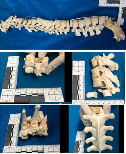
Figure 9. The sacrum and left ilium showed chop wounds. Cut marks, fracture lines, false starts, and hard tissue loss were observed on these bone surfaces.
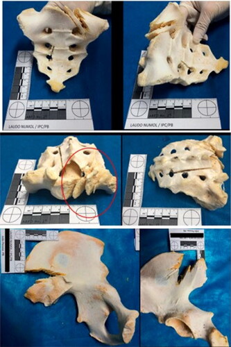
Figure 10. Blunt, sharp, and chop injuries at the distal epiphysis of the femurs and their diaphysis.
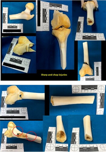
Figure 11. Distal epiphysis of the left tibia showed injuries consistent with false starts.