Figures & data
Figure 1. Posterior view of the human carbonized remains surrounded by multiple metallic filaments that involve the cervical and thoracoabdominal regions (A). High-density filaments in the radiological examination (B) and a close view of the filaments after opening the body bag (C).
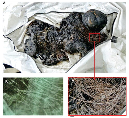
Figure 2. Radiological confrontation posing some of the elements used for this positive identification: morphology of the sella turcica (red arrows) and the aspect of the axis bone trabeculate (yellow ellipse). (A) Antemortem examination in 2009 provided by family members of the missing person. (B) Postmortem examination of the unidentified body performed by the forensic team in 2014.
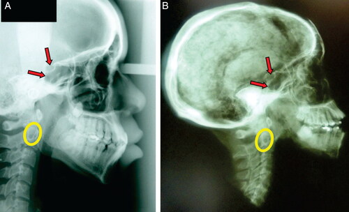
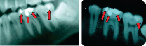
Figure 3. Periapical radiological confrontation showing some of the elements used for the positive identification: anatomy, angulation, and proportion of the first and second lower molars; the location, morphology, and proportion of dental treatments (red arrows) of the first and second lower molars. (A) Antemortem examination in 2009 provided by family members of the missing person. (B) Postmortem examination of the unidentified body performed by the forensic team in 2014.