Figures & data
Figure 1. Micrographs of fungi associated with southern right whale neonate skin. (A, B) Scanning electron micrographs showing fungal growth on the whale skin. (B) Scanning electron micrograph of an early formation of a fruiting body. (C) Fruiting bodies (ascomata) of C. globosum on a section of whale skin. (D) Light micrograph of unusually formed cells of C. globosum starting to form the textura intrica/epidermoidea of the peridium (fruit body wall).
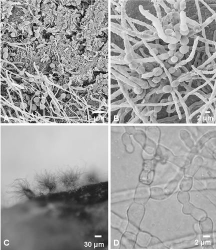
Table 1. Fungal isolates taken from different skin sections of a southern right whale neonate, cultured on different growth media, identified using morphology and sequencing data from the 26S and ITS regions of the ribosomal gene cluster
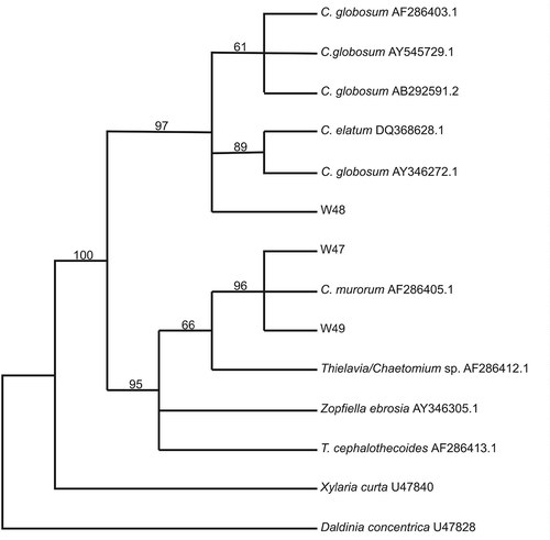
Figure 2. Maximum parsimony phylogenetic tree based on rDNA partial large subunit sequences (±500 bp) of whale skin fungal isolates and selected sequences from Genbank. Numbers above branches indicate bootstrap values (>50%) from 1000 replicates.