Phylogeny and intercontinental distribution of the pneumocandin-producing anamorphic fungus Glarea lozoyensis
F. Peláez
Spanish National Cancer Research Center, Melchor Fernández Almagro 3, Madrid, 28029, Spain
,
J. Collado
Oficina Española de Patentes y Marcas, Departamento de Patentes e Información Tecnológica, Paseo de la Castellana 75, Madrid, E-28071, Spain
,
G. Platas
Fundación MEDINA, Centro de Excelencia en Investigación de Medicamentos Innovadores en Andalucía, Parque Tecnológico de Ciencias de la Salud, Avda. de Conocimiento 3, E-18100, Armilla, Granada, Spain
,
D.P. Overy
University of Prince Edward Island, Duffy Research Center (NRC-INH), 550 University Avenue, Charlottetown, Prince Edward Island, C1A 4P3, Canada
,
J. Martín
Fundación MEDINA, Centro de Excelencia en Investigación de Medicamentos Innovadores en Andalucía, Parque Tecnológico de Ciencias de la Salud, Avda. de Conocimiento 3, E-18100, Armilla, Granada, Spain
,
F. Vicente
Fundación MEDINA, Centro de Excelencia en Investigación de Medicamentos Innovadores en Andalucía, Parque Tecnológico de Ciencias de la Salud, Avda. de Conocimiento 3, E-18100, Armilla, Granada, Spain
,
A. González del Val
Centro de Investigación Básica, Merck, Sharp and Dohme de España, S.A. Josefa Valcárcel 38, Madrid, E-28026, Spain
,
A. Basilio
Centro de Investigación Básica, Merck, Sharp and Dohme de España, S.A. Josefa Valcárcel 38, Madrid, E-28026, Spain
,
M. De la Cruz
Fundación MEDINA, Centro de Excelencia en Investigación de Medicamentos Innovadores en Andalucía, Parque Tecnológico de Ciencias de la Salud, Avda. de Conocimiento 3, E-18100, Armilla, Granada, Spain
,
J.R. Tormo
Fundación MEDINA, Centro de Excelencia en Investigación de Medicamentos Innovadores en Andalucía, Parque Tecnológico de Ciencias de la Salud, Avda. de Conocimiento 3, E-18100, Armilla, Granada, Spain
,
A. Fillola
Centro de Investigación Básica, Merck, Sharp and Dohme de España, S.A. Josefa Valcárcel 38, Madrid, E-28026, Spain
,
F. Arenal
PharmaMar S.A.U., Microbiology Department, R and D Drug Discovery, Edificio Parque Científico de Madrid, Santiago Grisolía 2, PTM, Tres Cantos, Madrid, E-28760, Spain
,
M. Villareal
Centro de Ciencias Medioambientales, CSIC, Serrano 115-bis, 28006, Madrid, Spain
,
V. Rubio
Centro de Ciencias Medioambientales, CSIC, Serrano 115-bis, 28006, Madrid, Spain
R. Galán
Departamento de Biología Vegetal, Facultad de Biología, Universidad de Alcalá, Alcalá de Henares, Madrid, E-28871, Spain
&
G.F. Bills
Fundación MEDINA, Centro de Excelencia en Investigación de Medicamentos Innovadores en Andalucía, Parque Tecnológico de Ciencias de la Salud, Avda. de Conocimiento 3, E-18100, Armilla, Granada, SpainCorrespondence[email protected]
Table 1. Strains and sequences. GenBank entries with FJ accession codes are new sequences obtained during this work; those with DQ codes are from Baral et al. (2006).
Table 2. Antimicrobial activity of four Glarea lozoyensis strains. The production media where some activity was detected is indicated in parentheses in each case. Codes for agar diffusion assays: A (clear inhibition zone >7 mm); B (clear inhibition zone < 7 mm or hazy inhibition zone >10 mm). See Vicente et al. (Citation2009) for media formulations.
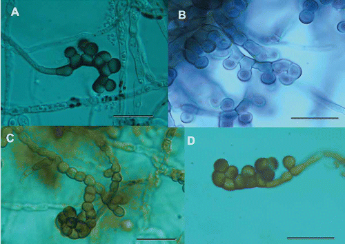
Figure 1. Developing conidiophores and conidia from four strains of Glarea lozoyensis. A. ATCC 20868=F-160870. B. F-226836. C. F-226838. D. F-239379. Scale bar = 20 μm.
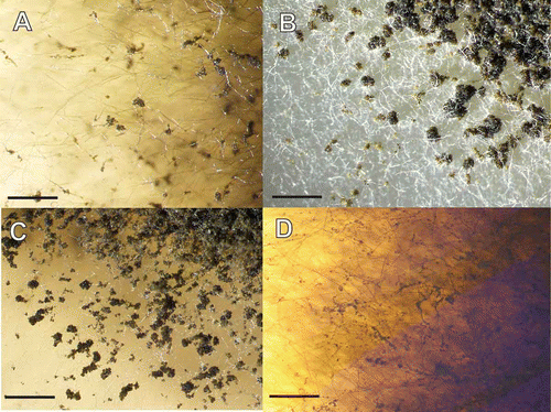
Figure 2. Accumulation of conidial masses on aerial hyphae of Glarea lozoyensis. Strains were grown on YM medium one month at room temperature. A. ATCC 20868=F-160870. B. F-226836. C. F-226838. D. F-239379. Scale bar = 2 mm.
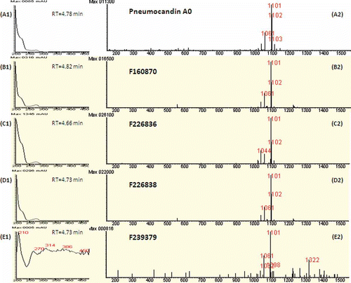
Figure 3. Identification of pneumocandin A0 in fermentation extracts of strains of Glarea lozoyensis. A1. UV spectrum of pneumocandin A0 with retention time of elution (4.78 min). A2. Positive ion mass spectrum of pneumocandin A0 (MW=1078 Da) at the same retention time. The combination of A1and A2 produce the fingerprint of pneumocandin A0. See methods for LC–MS protocols. B1, C1, D1 and E1 are the UV spectra of extracts of the four strains at the corresponding retention time, and B2, C2, D2 and E2 are the positive ion mass spectra of the respective sample at the same retention time as for the UV spectrum. All UV spectra were similar to authentic pneumocandin A0, except for E1 where the signal was near baseline. Similarly, all the positive ion mass spectra indicated pneumocandin A0 was in all samples.
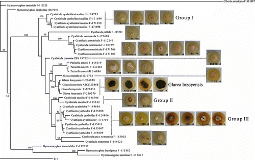
Figure 4. Phylogenetic tree of Glarea lozoyensis and related species generated by Bayesian analysis of combined ITS and 28S rRNA partial sequences. Ciboria americana was designated the outgroup. Clade credibility values are indicated at the branches. Colony morphology of G. lozoyensis and Cyathicula strains are mapped onto their corresponding branches.
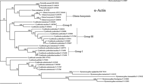
Figure 5. Phylogenetic tree of Glarea lozoyensis and related species generated by Bayesian analysis of α-actin gene partial sequences. Clade credibility values are indicated at the branches.
Table 3. Antimicrobial activity of Cyathicula and Pezizella strains against a panel of bacteria and fungi. The media where activity was detected are indicated in parentheses in each case. Media formulations have been previously reported (Vicente et al. Citation2009). Codes for agar diffusion assays: A (clear inhibition zone >7 mm); B (clear inhibition zone < 7 mm or hazy inhibition zone >10 mm); C (hazy inhibition zone < 10 mm).
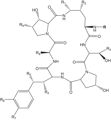
Figure 6. Structure of the echinocandins showing the common features and the radicals varying across the different molecular subtypes. Refer to for R-group substitutions.
Table 4. Naturally occurring echinocandins and producing organisms.
Benz
,
F
,
Knüsel
,
F
,
Nüesc
,
J
,
Treichler
,
H
,
Voser
,
W
,
Nyfeler
,
R
and
Keller-Schierlein
,
W
.
1974
.
Stoffwechselprodukte von Mikroorganismen. Echinocandin B, ein neuartiges Polypetid-Antibioticum aus Aspergillus nidulans var. echinulatus: Isolierung und Bausteine
.
Helv Chim Acta
,
57
:
2459
–
2477
.
Keller-Juslén
,
C
,
Kuhn
,
M
,
Loosli
,
HR
,
Petcher
,
TJ
,
Weber
,
HP
and
von Wartburg
,
A
.
1976
.
Struktur des cyclopeptide –antibiotikums SL 7810 (=Echinochandin B)
.
Tetrahedron Lett.
,
46
:
4147
–
4150
.
Traber
,
R
,
Keller-Juslén
,
C
,
Loosli
,
H-R
,
Kuhn
,
M
and
von Wartburg
,
A
.
1979
.
Cyclopeptid-Antibiotika aus Aspergillus-Arten
.
Struktur der Echinocandin C und D. Helv Chim Acta
,
62
:
1252
–
1267
.
Mizuno
,
K
,
Yagi
,
A
,
Satoi
,
S
,
Takada
,
M
,
Hayashi
,
M
,
Asano
,
K
and
Matsuda
,
T
.
1977
.
Studies on aculeacin. I. Isolation and characterization of aculeacin A
.
J Antibiot.
,
30
:
297
–
302
.
Satoi
,
S
,
Yagi
,
A
,
Asano
,
K
,
Mizuno
,
K
and
Watanabe
,
T
.
1977
.
Studies on aculeacin. II. Isolation and characterization of aculeacins B, C, D, E, F and G
.
J Antibiot.
,
30
:
303
–
307
.
Schwartz
,
RE
,
Giacobbe
,
RA
,
Bland
,
JA
and
Monaghan
,
RL
.
1989
.
L-671,329, a new antifungal agent. I. Fermentation and isolation
.
J Antibiot.
,
42
:
163
–
167
.
Bills
,
GF
,
Martín
,
J
,
Collado
,
J
,
Platas
,
G
,
Overy
,
D
,
Tormo
,
JR
,
Vicente
,
F
,
Verkley
,
G
and
Crous
,
PW
.
2009
.
Measuring the distribution and diversity of antibiosis and secondary metabolites in the filamentous fungi
.
Soc Ind Microbiol News
,
59
:
133
–
146
.
Schwartz
,
RE
,
Sesin
,
DF
,
Joshua
,
H
,
Wilson
,
KE
,
Kempf
,
AJ
,
Goklen
,
KA
,
Kuehner
,
D
,
Gailliot
,
P
,
Gleason
,
C
,
White
,
R
,
Inamine
,
E
,
Bills
,
G
,
Samon
,
P
and
Zitano
,
L
.
1992
.
Pneumocandins from Zalerion arboricola. I. Discovery and isolation
.
J Antibiot.
,
45
:
1853
–
1866
.
Petersen
,
LA
,
Hughes
,
DL
,
Hughes
,
R
,
Di Michele
,
L
,
Salmon
,
P
and
Connors
,
N
.
2001
.
Effects of amino acid and trace element supplementation on pneumocandin production by Glarea lozoyensis: impact on titer, analogue levels, and the identification of new analogues of pneumocandin B0
.
J Ind Microbiol Biotechnol.
,
26
:
216
–
221
.
Hino
,
M
,
Fujie
,
A
,
Iwamoto
,
T
,
Hori
,
Y
,
Hashimoto
,
M
,
Tsurumi
,
Y
,
Sakamoto
,
K
,
Takase
,
S
and
Hashimoto
,
S
.
2001
.
Chemical diversity in lipopeptide antifungal antibiotics
.
J Ind Microbiol Biotechnol.
,
27
:
157
–
162
.
Kanasaki
,
R
,
Abe
,
F
,
Kobayashi
,
M
,
Katsuoka
,
M
,
Hashimoto
,
M
,
Takase
,
S
,
Tsurumi
,
Y
,
Fujie
,
A
,
Hino
,
M
,
Hashimoto
,
S
and
Hori
,
Y
.
2006b
.
FR220897 and FR22089, novel antifungal lipopeptides from Coelophoma empetri No. 14573
.
J Antibiot.
,
59
:
149
–
157
.
Kanasaki
,
R
,
Kobayashi
,
M
,
Fujine
,
K
,
Sato
,
I
,
Hashimoto
,
M
,
Takase
,
S
,
Tsurumi
,
Y
,
Fujie
,
A
,
Hino
,
M
,
Hashimoto
,
S
and
Hori
,
Y
.
2006c
.
FR227673 and FR190293, novel antifungal lipopeptides from Chalara sp. No. 22210 and Tolypocladium parasiticum No. 16616
.
J Antibiot.
,
59
:
158
–
167
.
Kanasaki
,
R
,
Sakamoto
,
K
,
Hashimoto
,
M
,
Takase
,
S
,
Tsurumi
,
Y
,
Fujie
,
A
,
Hino
,
M
,
Hashimoto
,
S
and
Hori
,
Y
.
2006a
.
FR209602 and related compounds, novel antifungal lipopeptides from Coleophoma crateriformis no.738 - I. Taxonomy, fermentation, isolation and physico-chemical properties
.
J Antibiot.
,
59
:
137
–
144
.
If you are unable to obtain permissions via Rightslink, please complete and submit this Permissions form. For more information, please visit our Permissions help page.