Figures & data
Figure 1. Secondary metabolites from Hamigera sp.
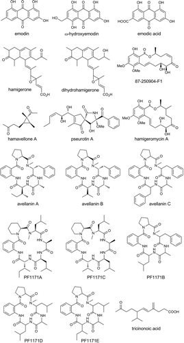
Table 1. Strains of the Hamigera clade examined for secondary metabolites.
Figure 2. HPLC chromatogram of culture extract of H. insecticola NRRL35442 (shaking culture, A-16 medium, 4 days, HPLC condition A monitored at 254 nm).
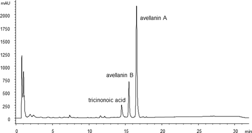
Figure 3. HPLC chromatogram of culture extract of H. insecticola NRRL35442 (static culture, A-16 medium, 21 days, HPLC condition A monitored at 254 nm).
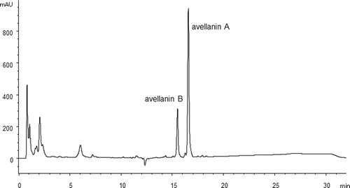
Table 2. Distribution of tricinonoic acid and peptide metabolites in Hamigera species.
Figure 4. HPLC chromatogram of culture extract of H. paravellanea NRRL35714 (shaking culture, supermalt, 4 days, HPLC condition B monitored at 254 nm).

Figure 5. HPLC chromatogram of culture extract of H. ingelheimensis NRRL29060 (shaking culture, A-3M, 4 days, HPLC condition B monitored at 254 nm).
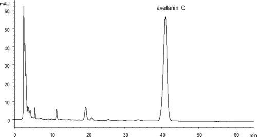
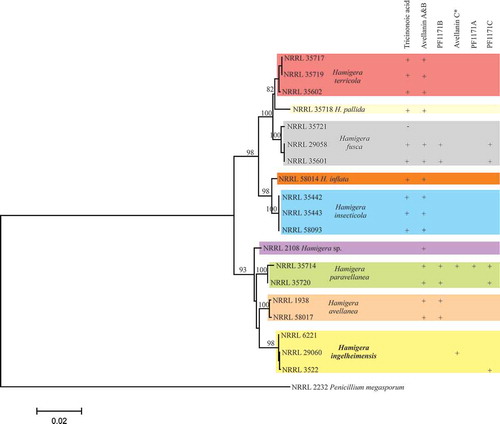
Figure 6. Phylogenetic tree showing the relations of Hamigera species.