Publication Cover
Mycology
An International Journal on Fungal Biology
Volume 10, 2019 - Issue 3
1,252
Views
6
CrossRef citations to date
0
Altmetric
Article

Quasipucciniastrum agrimoniae, gen. et sp. nov. on Agrimonia (Rosaceae) from China

, &
Pages 141-150 | Received 31 Mar 2019, Accepted 17 Apr 2019, Published online: 30 Apr 2019

Figures & data

Table 1. Herbarium specimens used for molecular phylogenetic analyses.

Table 2. Sequence data retrieved from GenBank and used for phylogenetic analyses.
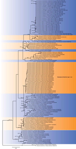
Figure 1. Bayesian 50% majority-rule consensus tree based on concatenated data of rDNA ITS and LSU sequences. Pileolaria terebinthi was used as outgroup. Values on the branches indicate maximum likelihood bootstrap values and Bayesian posterior probabilities. Hyphen indicates that bootstrap values were less than 75% and Bayesian posterior probabilities less than 0.80.

Figure 1. Bayesian 50% majority-rule consensus tree based on concatenated data of rDNA ITS and LSU sequences. Pileolaria terebinthi was used as outgroup. Values on the branches indicate maximum likelihood bootstrap values and Bayesian posterior probabilities. Hyphen indicates that bootstrap values were less than 75% and Bayesian posterior probabilities less than 0.80.

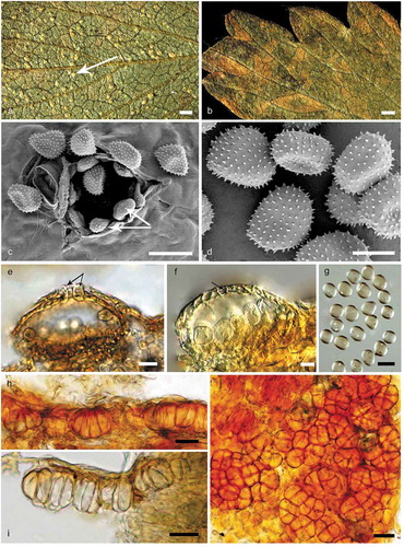
Figure 2. Morphological characters of Quasipucciniastrum agrimoniae. A: Uredinia (white arrow) on the hypophyllous leaf surface. B: No uredinia and telia on eiphyllous leaf surface. C: Uredinium with echinulate ostiolar cells (white arrow) observed by SEM. D: Urediniospores with echinulate spines observed by SEM. E: Uredinium with well-developed ostiolar cells (black arrow) observed by LM. F: Uredinium with peridium cell (black arrow). G: Urediniospores observed by LM. H: Vertical section of hypophyllous telia. I: Subepidermal teliospores with apparently thickened apical wall. J: Subglobose teliospores with vertical or oblique septa. Bars: A, B = 0.6 mm; C, E, G, H, I, J = 20µm; D, F = 10µm.

Figure 2. Morphological characters of Quasipucciniastrum agrimoniae. A: Uredinia (white arrow) on the hypophyllous leaf surface. B: No uredinia and telia on eiphyllous leaf surface. C: Uredinium with echinulate ostiolar cells (white arrow) observed by SEM. D: Urediniospores with echinulate spines observed by SEM. E: Uredinium with well-developed ostiolar cells (black arrow) observed by LM. F: Uredinium with peridium cell (black arrow). G: Urediniospores observed by LM. H: Vertical section of hypophyllous telia. I: Subepidermal teliospores with apparently thickened apical wall. J: Subglobose teliospores with vertical or oblique septa. Bars: A, B = 0.6 mm; C, E, G, H, I, J = 20µm; D, F = 10µm.