Figures & data
Table 1. Averaged daily colony radial growth and standard deviation, in mm, on Rye B agar. Three plates were used for each temperature, and the experiment was repeated three times.
Figure 1. Pythiogeton manoomin. (a), Colony on PDA; (b–c), hyphae and appressoria (arrows) on RSA; (d–e), sporangia, terminal or intercalary and short distance from the end of supporting hyphae; (f–g), globose (arrow) and ellipsoid sporangia, (h–i), reniform sporangia, (j), bilobate sporangia, (k–l), pyriform sporangia, (m–n), globose (arrow) and ovoid sporangia; (o), chlamydospores, (p–q), sporangia initiating formation of the discharge tube (arrowheads), (r), formation of zoospores from a protoplasm mass, and, (s), discharged zoospores; (t), empty sporangia, showing empty discharge tubes (arrows). Scale bars = 20 μM.
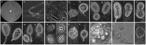
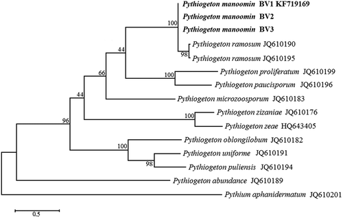
Figure 2. The Maximum Parsimony tree of Pythiogeton species with Pythium aphanidermatum as the outgroup based on ITS sequences. Numbers on the branches represent bootstrap values obtained from 1000 replications.