Figures & data
Figure 1. CFUs detected from tomato leaflets inoculated with a GFP-expressing strain Amgfp derived from Akanthomyces muscarius IMI 268317. (a) Total CFUs detected from leaf disk inoculated with a conidial suspension drop. (b) Proportion of CFUs detected from washed leaf disk.
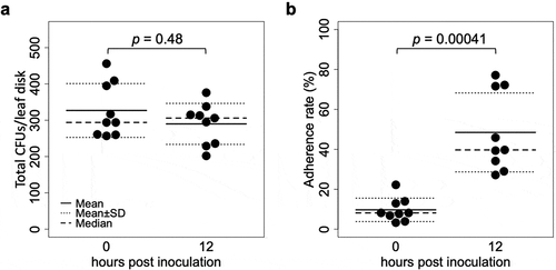
Figure 2. The number of colonies forming units over time detected on tomato leaflets treated with conidial suspension of 1 × 10⁵ (a) or 1 × 10⁶ (b) conidia/ml of a GFP-expressing strain Amgfp derived from Akanthomyces muscarius IMI 268317. Lines indicate medians.
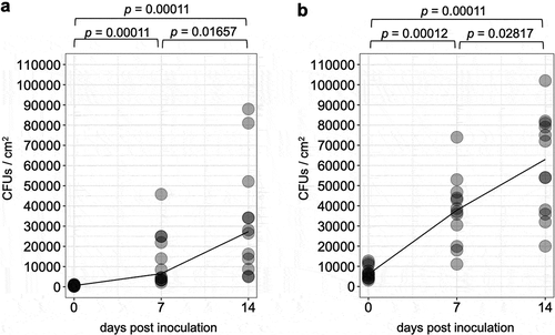
Figure 3. Laser scanning confocal microscopy images of epiphytic colonisation of a GFP-expressing strain Amgfp derived from Akanthomyces muscarius IMI 268317 on tomato leaflets. Viable conidia and hyphae were detected on tomato leaflets surfaces (a–d) and cross section (e) at 1 dpi (a), 3 dpi (b and e), 7 dpi (c), and 14 dpi (d). Microcycle conidiation like structures in (b) and phialides with conidia (c and d) were arrowed. Scale bar = 100 µm.
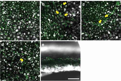
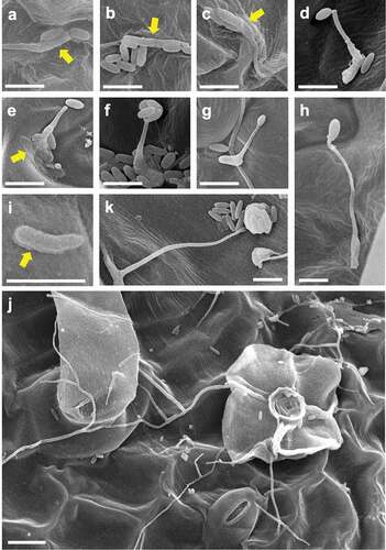
Figure 4. Scanned electron microscopy images of epiphytic colonisation of a GFP-expressing strain Amgfp derived from Akanthomyces muscarius IMI 268317 on tomato leaflets at 1 dpi (a–c), 3 dpi (d, f–i), and 7 dpi (e, j, k). (a–c) ambiguated boundaries between germinated conidia and adjacent conidia or leaf surface, (d) a germ-tube with a conidium on tip, (e) a germ-tube with a conidium adjacent to a tip, and mucilage-like materials around a conidium (arrow), (f) multiple conidia on germ-tube tip, (g) a germ-tube with a swollen tip, (h) a germ-tube with an appressorium-like swollen tip, (i) a germinated conidia with mucilage-like materials (arrow), (j) conidia and elongated hyphae around a trichome, (k) a mass of conidia produced on a tip of a solitary phialide. Scale bar, 5 µm (a–i, k), 10 µm (j).