Figures & data
Table 1. Median inhibitory concentration (IC50) values of the methanolic extract of Padina tetrastromatica in free radical scavenging assays.
Figure 1. Percentage of inhibition of MCF-7 cells versus different concentrations of the methanolic extract of Padina tetrastromatica. MCF-7 cells were treated with various concentrations of the extract (100, 200, 300, 400 and 500 µg/ml) or 0.1% dimethyl sulfoxide (DMSO; vehicle control) for 48 h. Cell viability of MCF-7 cells was determined by the 3-(4,5-dimethylthiazol-2-yl)-2,5-diphenyltetrazolium bromide (MTT) assay.
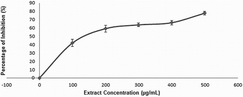
Figure 2. Comet tail length of the extract-treated 3T3-L1 and MCF-7 cells relative to hydrogen peroxide (H2O2)-treated and untreated negative controls. 3T3-L1 and MCF-7 cells were treated with various concentrations of the extract (5, 10, 15, 20 and 25 µg/ml) or 0.2 µg/ml of doxorubicin, as the positive control. Untreated cells and cells treated with 100 mM of H2O2 served as negative controls. Data are shown as mean ± SD. *p < 0.05, **p < 0.01.
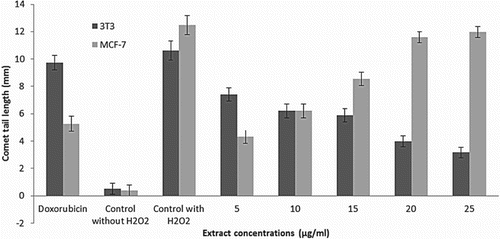
Figure 3. Percentage of DNA damage in extract-treated 3T3-L1 and MCF-7 cells relative to hydrogen peroxide (H2O2)-treated and untreated negative controls. 3T3-L1 and MCF-7 cells were treated with various concentrations of the extract (5, 10, 15, 20 and 25 µg/ml) or 0.2 µg/ml of doxorubicin, as the positive control. Untreated cells and cells treated with 100 mM of H2O2 served as negative controls. Data are shown as mean ± SD. *p < 0.05, **p < 0.01.
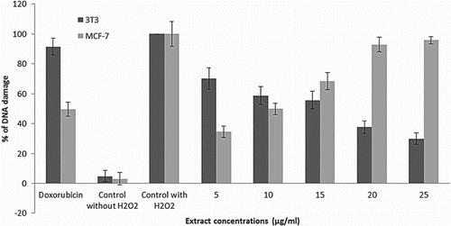
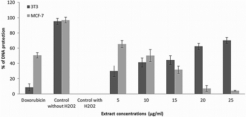
Figure 4. Percentage of DNA protection in extract-treated 3T3-L1 and MCF-7 cells relative to hydrogen peroxide (H2O2)-treated and untreated negative controls. 3T3-L1 and MCF-7 cells were treated with various concentrations of the extract (5, 10, 15, 20 and 25 µg/ml) or 0.2 µg/ml of doxorubicin, as the positive control. Untreated cells and cells treated with 100 mM of H2O2 served as negative controls. Data are shown as mean ± SD. *p < 0.05, **p < 0.01.