Figures & data
Figure 1. Light microscope view of a seminiferous tubule in the Pelamis platarus testis. The seminiferous tubule has a wide lumen and thick germinal epithelium containing developing germ cells (Spermatogonia & Spermatocytes, Spermatids). Bar = 50 µm.
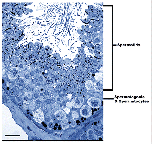
Figure 2. Early round spermatids undergoing acrosome development during spermiogenesis within the seminiferous epithelium of Pelamis platarus. (A) The acrosomal vesicle (Av) is juxtapositioned to the apical portion of the nucleus (Nu). The vesicle is in the early phase of growth and contains an acrosomal granule (black arrow). The cytoplasm of the spermatid has numerous mitochondria (Mi) and a prominent nuage (Ng). Bar = 2 µm. (B) Sagittal view of an acrosome later in development showing the attachment of the acrosomal vesicle (Av) to the apical portion of the nucleus (Nu) and the presence of the acrosomal granule (black arrow). Bar = 2 µm. (C) The caudal portion of the nucleus houses the proximal centriole (Pc) attached to the growing distal centriole/neck (Dc). Pp, principal piece; Pm, pericentriolar material. Bar = 1 µm. (D) The Golgi apparatus is prominent near the apex of the nucleus in round spermatids. Transport vesicles (black arrows) can be seen budding off of proximal cisterna of the Golgi and presumably will merge with the acrosome during its growth phase. Bar = 0.5 µm.
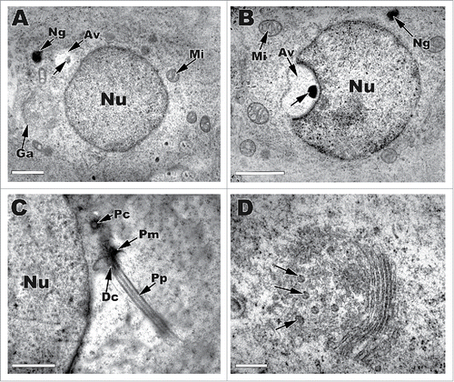
Figure 3. Late stages of round spermatid development in Pelamis platarus. (A) The acrosomal vesicle (Av) has made a deep indentation into the apical nucleus (Nu). The acrosomal granule (black arrow) is now located basally within the acrosome. A prominent subacrosomal space (Sa) develops under the acrosomal vesicle. (B,C) The centrioles (Pc, Dc), which are housed in the caudal nuclear fossa (Nf) continue to contribute to the growing principal piece (Pp). Mitochondria (Mi) start to relocate to the area next to the caudal distal centriole. Nu, nucleus; Sa, subacrosomal space; Av, acrosomal vesicle; black arrow, acrosomal granule; white arrow, subascrosomal granule. (D) At the end of the round spermatid stage, the caudal end of the nucleus begins to elongate and chromatin condenses in a fibrous fashion (Nu). The acrosomal granule (black arrow) has reached its maximum size within an acrosomal vesicle (Av) that has collapsed on to the apical nuclear surface. Sa, subascrosomal space. Bar = 2 µm for all micrographs.
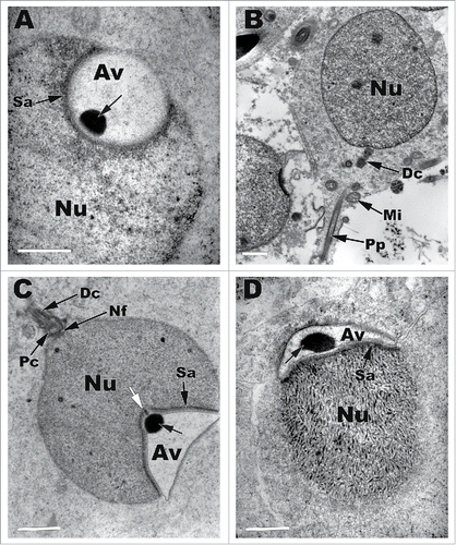
Figure 4. The middle stage of spermatid elongation within the seminiferous epithelium of Pelamis platarus. (A) The acrosomal vesicle (Av) has flattened and starts to envelop the elongating nucleus (Nu). The chromatin within the apical nucleus shows spiraling and large open nucleoplasmic spaces (white arrow) form. Sa, Subacrosomal space; Ns, nuclear shoulders. Bar = 2 µm. (B) The chromatin is shown condensing in a spiral fashion (*) with nucleoplasmic spaces (white arrow) abundant within the nucleus in CS. Bar = 2 µm. (C) Caudal end of nucleus shows the 2 shoulders (Ns), which are devoid of chromatin and can be seen lateral to the insert of the flagellum within the flagellar/nuclear fossa (Nf). Bar = 1 µm. (D) The acrosomal complex under high power. Av, acrosomal vesicle; Ag, acrosomal granule; Ar, acrosomal lucent zone; Nr, nuclear rostrum; Sa, subacrosomal space, large black arrowheads, desmosomes; small single black arrowhead, epinuclear lucent zone. Bar = 1 µm.
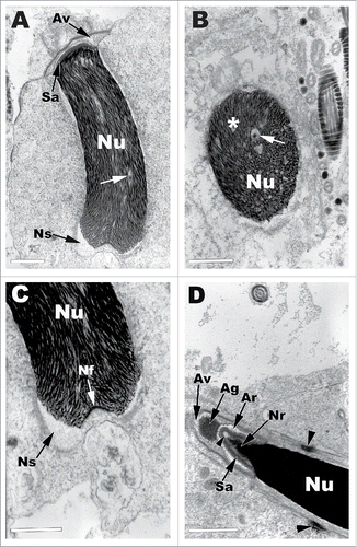
Figure 5. An elongating spermatid nearing the climax of elongation in the seminiferous epithelium of Pelamis platurus. The acrosomal complex and apical nucleus is on display in sagittal section (A,E) and with represented cross sections through the letters B,C,D,F,G. All of the major parts of this complex that are found in the typical ophidian spermatozoa are present. Ac, acrosomal complex; Ma, manchette; Av, acrosomal vesicle; black arrowhead and Pe, perforatorium; Sa, subascrosomal space; Ar, acrosomal lucent zone; Sm, Sertoli cell membrane laminae; white arrow and EP, epinuclear lucent zone; Nr, nuclear rostrum; Bp, basal plate; white arrowhead, nuclear lacuna. Bars = 1 µm.
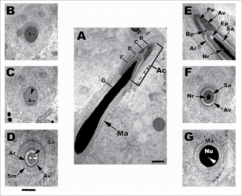
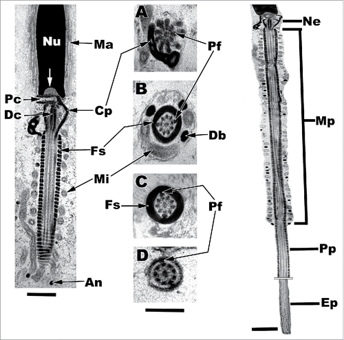
Figure 6. Sagittal and cross sections of the caudal nucleus and components of the flagellum of a late staged elongate in the Pelamis platurus testis. Cross sections: A, distal centriole; B, midpiece; C, principal piece; D, endpiece. Ne, neck; Mp, midpiece; Pp, principal piece; Ep, endpiece; Nu, nucleus; Ma, manchette; Pc, proximal centriole; Dc, distal centriole; Cp, dense collar; Fs, fibrous sheath; Mi, mitochondria; An, annulus; Pf, peripheral fibers; Db, dense bodies; white arrow, nuclear fossa. Bar = 1 µm.