389
Views
0
CrossRef citations to date
0
Altmetric
Articles

Relationship between initiation of embryogenic suspensor mass (ESM) and zygotic embryo development in Pinus densiflora

&
Pages 166-171 | Received 04 Dec 2013, Accepted 28 Jan 2014, Published online: 26 Feb 2014

Figures & data

Table 1. Collection locations, dates and developmental stages of embryos used for ESM initiation in P. densiflora.

Figure 1. Developmental embryo stages of P. densiflora. (a) Archegonia (arrows) from a 31 May seed. (b) No embryos found in 7 June seeds. (c) Early-stage embryos in corrosion cavity (13 June). (d) Magnified picture of Figure 1c. One small globular zygotic embryo in corrosion cavity, e: embryonal head, s: suspensor, m: micropyle. (e) Proembryos in the corrosion cavity (cc), s: suspensor (21 June). (f) Numerous suspensors in the corrosion cavity (magnified Figure 1e). (g) Late-stage proembryos in corrosion cavity (28 June). (h) Embryonal head of each embryo (arrow) has more divided cells and longer suspensors than shown in those of Figure g. (i) One surviving dominant embryo (arrow); the other embryo has degenerated (5 July). (j) Magnified picture of Figure c. (k) A zygotic embryo developed more vigorously (13 July). (l) Cotyledon-stage embryo in the corrosion cavity (20 July). Bars: 420 μm (a–d), 179 μm (e), 159 μm (i), 140 μm (j), 117 μm (k), and 147 μm (l).
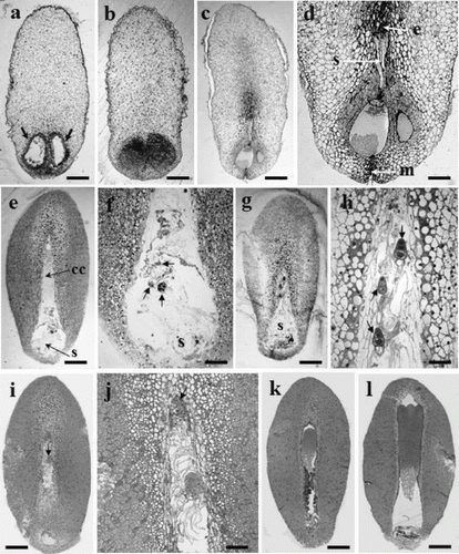
Figure 1. Developmental embryo stages of P. densiflora. (a) Archegonia (arrows) from a 31 May seed. (b) No embryos found in 7 June seeds. (c) Early-stage embryos in corrosion cavity (13 June). (d) Magnified picture of Figure 1c. One small globular zygotic embryo in corrosion cavity, e: embryonal head, s: suspensor, m: micropyle. (e) Proembryos in the corrosion cavity (cc), s: suspensor (21 June). (f) Numerous suspensors in the corrosion cavity (magnified Figure 1e). (g) Late-stage proembryos in corrosion cavity (28 June). (h) Embryonal head of each embryo (arrow) has more divided cells and longer suspensors than shown in those of Figure g. (i) One surviving dominant embryo (arrow); the other embryo has degenerated (5 July). (j) Magnified picture of Figure c. (k) A zygotic embryo developed more vigorously (13 July). (l) Cotyledon-stage embryo in the corrosion cavity (20 July). Bars: 420 μm (a–d), 179 μm (e), 159 μm (i), 140 μm (j), 117 μm (k), and 147 μm (l).

Figure 2. Mucilaginous ESM extruded from the micropyle end of megagametophyte after 8 weeks in culture. Bar: 8 mm.
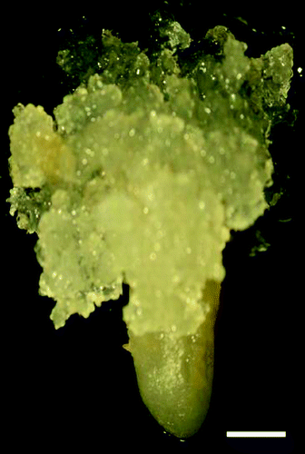
Figure 2. Mucilaginous ESM extruded from the micropyle end of megagametophyte after 8 weeks in culture. Bar: 8 mm.

Figure 3. Sections of immature P. densiflora embryos correlated with collection dates and locations. (a) Two embryos on the left (28 June 2005, Suwon), two in middle (1 July 2005, Suwon), two on the right (5 July 2005, Suwon). (b) Two embryos on left (28 June 2005, Anmyeondo), two in middle (1 July 2005, Anmyeondo), two on right (5 July 2005, Anmyeondo) (c) two embryo on left (28 June 2006, Suwon), two in middle (1 July 2006, Suwon), two on right (1 July 2006, Suwon). Bar: 1.2 mm (a–c).
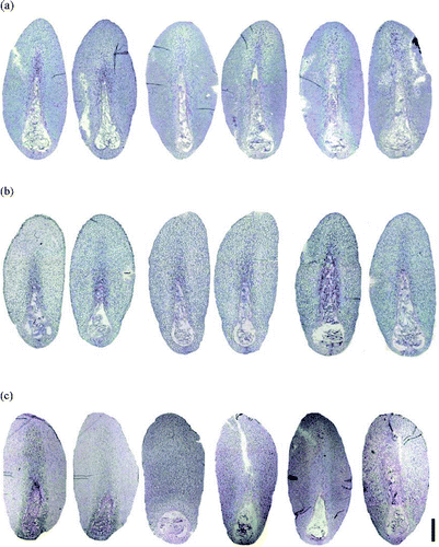
Figure 3. Sections of immature P. densiflora embryos correlated with collection dates and locations. (a) Two embryos on the left (28 June 2005, Suwon), two in middle (1 July 2005, Suwon), two on the right (5 July 2005, Suwon). (b) Two embryos on left (28 June 2005, Anmyeondo), two in middle (1 July 2005, Anmyeondo), two on right (5 July 2005, Anmyeondo) (c) two embryo on left (28 June 2006, Suwon), two in middle (1 July 2006, Suwon), two on right (1 July 2006, Suwon). Bar: 1.2 mm (a–c).

Reprints and Corporate Permissions

Please note: Selecting permissions does not provide access to the full text of the article, please see our help page How do I view content?

To request a reprint or corporate permissions for this article, please click on the relevant link below:

Academic Permissions

Please note: Selecting permissions does not provide access to the full text of the article, please see our help page How do I view content?

Obtain permissions instantly via Rightslink by clicking on the button below:

If you are unable to obtain permissions via Rightslink, please complete and submit this Permissions form. For more information, please visit our Permissions help page.