Figures & data
Figure 1. Schema of the poly(lactic acid)-based carrier (A). Photographs of the surface of carrier in the presence or absence of the coating (B). The images were taken by using VHX-5000 digital microscope (Keyence Corp.). Original magnification, ×400. Bar; 100 μm. Comparison of coating-free and coated carrier thicknesses (C). The experiment was conducted 3 times and the mean ± SE is indicated. ** P< 0.01 by one-way ANOVA. A procedure of the carrier application (D). The carrier adhered to cell sheets after being overlaid on the sheets for approximately 5 min. After transfer of cell sheets to the affected sites, dropping of pre-warmed physiological solution onto the carrier allowed cell sheets detachment.
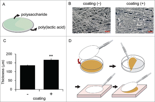
Figure 2. Comparison of the warp height between poly(lactic acid)-based carrier and Product A (A). The experiment was conducted 3 times and the mean ± SE is indicated. **P < 0.01 compared to Product A by one-way ANOVA. Representative data of warping in Product A (B). The arrow indicates the warp height.
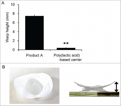
Figure 3. Comparison of the required time for cell sheets detachment in experimental transplantation of cell sheets onto the cornea (A). Each carrier along with cell sheets was applied onto the cornea, and pre-warmed saline (isotonic sodium chloride solution) was dropped from the upper part of each carrier for the indicated times and the duration required for cell sheet detachment was compared. The experiment was conducted 3 times and the mean ± SE is indicated. *P < 0.05, **P < 0.01, compared to Product A by one-way ANOVA. Representative image of cell sheet transplantation onto the surface of the canine cornea (B).
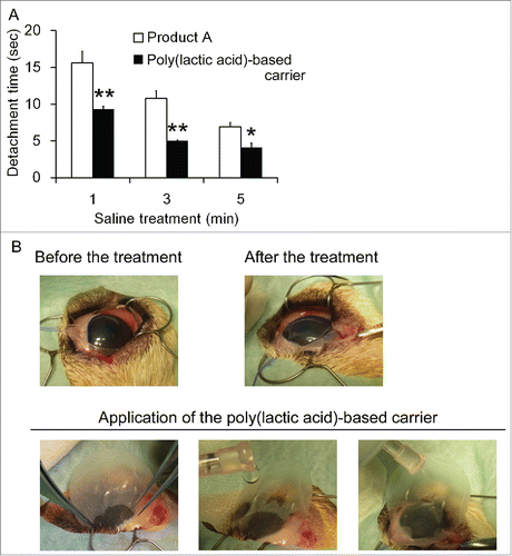
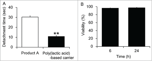
Figure 4. Comparison of the required time for cell detachment in experimental transplantation of cell sheets onto the rat dermis (A). Each carrier along with cell sheets was applied onto the dermis, and pre-warmed saline (isotonic sodium chloride solution) was dropped from the upper part of each carrier for 5 min and the duration required for cell sheet detachment was compared. The experiment was conducted 3 times and the mean ± SE is indicated. **P < 0.01 compared to Product A by one-way ANOVA. Cell viabilities of rat cells which was detached from the carrier (B). Rat cell sheets were bound to the carrier, treated with saline (isotonic sodium chloride solution) at 42°C for 5 min, cultured in the medium for indicated time and cell viabilities were determined. The experiment was conducted 3 times and the mean ± SE is indicated.