Figures & data
Figure 1. Model illustrating various membrane trafficking routes. This Figure explains basic membrane trafficking routes. (1) TGN exit mediated by AP-1A. (2) TGN exit mediated by AP-4. (3) TGN exit toward REs, regulated by Rab13. (4a) Apical sorting from REs. (4b) AP-1B-mediated sorting to the basolateral membrane. (5) Sorting into late endosomes/lysosomes. (6) Retrieval from late endosomes/lysosomes to the TGN. (7) Retrieval from BEEs to the TGN. (8a) Apical endocytosis and fast recycling from AEEs. (8b) Basolateral endocytosis and fast recycling from BEEs. (9a and 9b) Delivery of internalized cargos to REs. See main text for more details. Note that pathways leading from REs toward the TGN, AEEs, BEEs, and late endosomes are not shown.
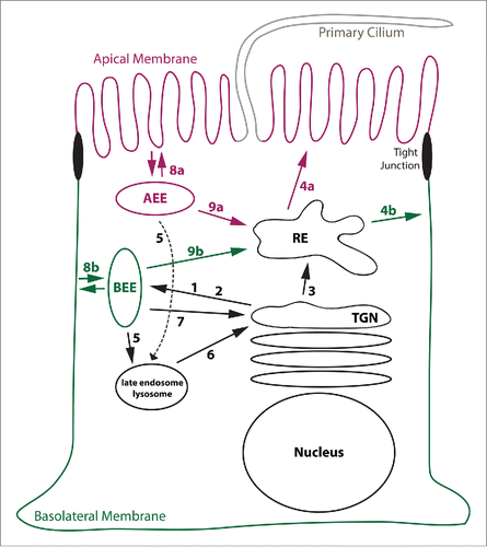
Table 1. Commonly used small protein tags
Figure 2. Model for Membrane recruitment of AP-1B. This figure represents a schematic drawing of the putative membrane recruitment of AP-1B indicating the rotation of the µ1B subunit upon membrane recruitment. Note that the C-terminus of µ subunits is organized in subdomains A and B as illustrated in the membrane-bound form. Whereas subdomain A binds to YxxØ sorting signals and thus cargo proteins, subdomain B probably binds to PtdIns(3,4,5)P3. Finally, Arf6 most likely binds to both γ and β1 adaptin. Abbreviations PI(3,4,5)P3, PtdIns(3,4,5)P3
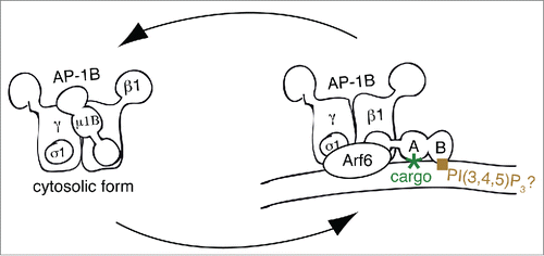
Table 2. Alignment of “loca” region in µ1 genes from various species (corresponding to residues 333 – 346 in µ1B)
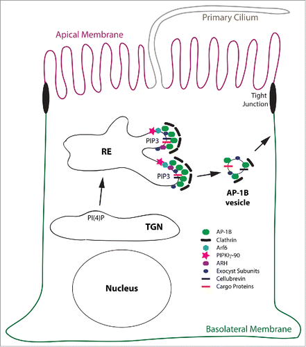
Figure 3. Model for the AP-1B domain in REs. This is a schematic depiction of the AP-1B domain in a RE leading to the generation of AP-1B vesicles that are targeted to the basolateral membrane. Proteins depicted are AP-1B, clathrin, Arf6, PIPKIγ-90, ARH, exocyst subunits, cellubrevin and cargo proteins. Note, that cellubrevin, exocyst subunits, and clathrin are confirmed components of AP-1B vesicles.Citation30,54 For simplicity reasons, the early secretory pathway including the Golgi apparatus, AEEs, BEEs, and other endocytic organelles as well as endocytic trafficking are not depicted. Abbreviation: PIP3, PtdIns(3,4,5)P3.