Figures & data
Figure 1. Surgical trauma induces adipose tissue browning. (A) Representative macroscopic morphology from paraffin embedded inguinal adipose tissues. From the left to the right: baseline, sham, incision and trauma group. (B) Representative H&E-stained inguinal adipose tissue sections which differentiated lipid droplets (white) from connective tissue (red). From the left to the right: baseline, sham, incision and trauma group. (C) Representative characterization of brown/beige adipocyte marker UCP1 by immunohistochemistry in inguinal (top), epididymal (middle), and interscapular (bottom) depots. Epididymal or white visceral and interscapular or brown fat represent negative and positive controls respectively. Data are representative of 10 independent animals per group. Bars represent 100 μm.
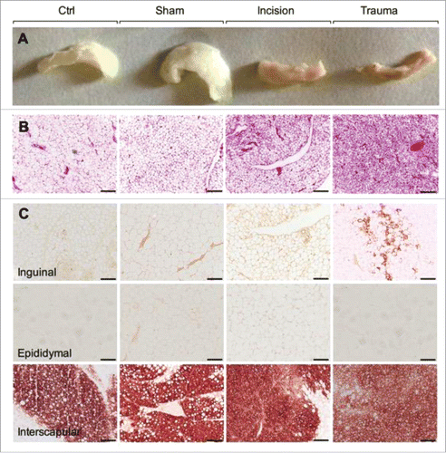
Figure 2. Trauma induces distant browning. (A, upper panel) Representative H&E stained inguinal fat from baseline and one day post trauma (site and contralateral). Trauma site and contralateral showed reduce lipid droplets to form packed multilocular adipocytes. (A, lower panel) Immunostaining using antibody against UCP1 showed protein expression in the traumatized (trauma Site) and contralateral adipose tissue. (B-D) Q-PCR against UCP1, UCP2, PGC1α, CD137 and TBX1 gene in the inguinal adipose depot (B) and against UCP1, UCP2 and Elovl3 gene in the interscapular (C) and perivascular fat depot (D) at baseline and one day after left inguinal trauma (trauma site and contralateral). Expression values were normalized to their respective baseline. Data are mean±SEM of 5–6 independent animals per group. Bars represent 20 μm. *p < 0.05, **p < 0.01, ***p < 0.001 vs. baseline.
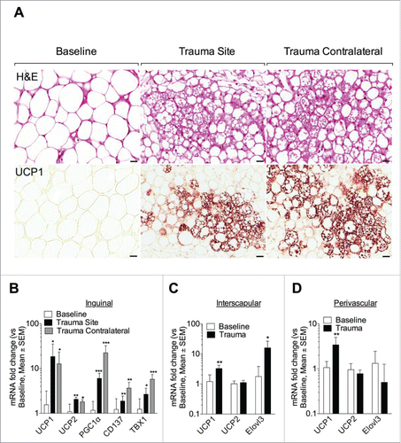
Figure 3. Trauma induces adipose browning up to day 5 following surgery. Representative H&E and UCP1-stained inguinal adipose tissue sections at different time points after surgical trauma as indicated. Data are representative of 4 independent animals per group. Bars represent 20 μm.
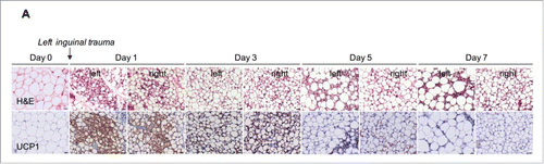
Figure 4. Short-term caloric restriction minimally modulates adipose browning relative to surgical trauma. (A, B) Caloric intake (A) and weight (B) were measured in animal fed with a NC or CR one day before diet randomization (Pre-CR, weight only), one, 2 and 3 days after (CR1, 2 and 3) and at one day post-op (Post-op). Caloric intake is expressed as Kcal/mouse/day and weight as gram. (C) Body temperature was continuously monitored during procedures (15 min) with a rectal probe. T1 (2 ∼ 3 min after the rectal probe insertion), T2 (15 min after the rectal probe insertion), and T3 (the highest temperature during measurement) and is expressed in degrees Celsius (°C). Data are mean±SEM of 6 independent animals per group. *p < 0.05 versus NC. (D, E) Animals were randomly exposed to a 3 day CR or NC before the indicated procedure (sham or trauma). (D) Q-PCR analysis of UCP1 in inguinal adipose tissue, at baseline or at one day (sham, trauma site and trauma contralateral). (E) Representative UCP1 immunohistochemistry in inguinal adipose tissue. Upper panel: NC group at baseline, sham, trauma site and contralateral (from left to the right). Lower panel: CR group at baseline, sham, trauma site and contralateral (from left to the right). Data are representative of 10 independent animals per group. Bars represent 100 μm. *p < 0.05 vs. baseline.
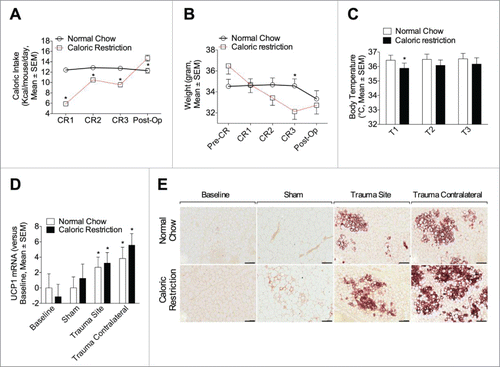
Figure 5. PDGFRα+ cells decrease after surgical injury. (A, B) Cells were isolated from inguinal adipose tissue at baseline and one day post left inguinal injury (trauma site) and analyzed for the expression of PDGFRα, CD34 (A) and Sca1 (B). The percentages of cells are indicated on the flow profile. The graphs show percentage of cells in inguinal depot at baseline and one day after left inguinal trauma (trauma site). Data are mean±SEM of 5–10 independent animals per group. *p < 0.05 versus baseline.
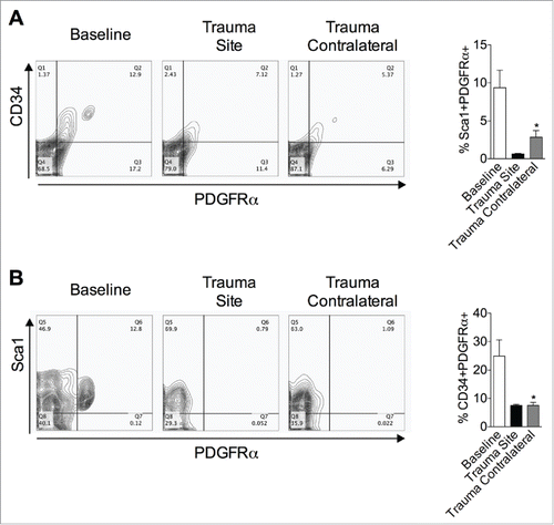
Figure 6. Surgical trauma promotes local monocyte recruitment and alternatively activated macrophages, but the remote browning can occur without increased macrophages in the browning adipose. (A-D) Cells were isolated from adipose tissue at baseline and one day post left inguinal injury (trauma site) and labeled for flow cytometry. (A) FACS analysis on cell surface marker expression of F4/80 and CD11b at baseline and one day after surgery. The graphs show percentage of F4/80+CD11b+ macrophages in inguinal depot at baseline and one day after left inguinal trauma (trauma site). (B-D) Macrophages were then assessed for the expression of CD11c (B), CD206 (C) and CD301 (D). Data are mean±SEM of 10 independent animals per group. *p < 0.05, **p < 0.01 vs. baseline. (E) Q-PCR against alternatively activated Arg1, Ym1, Fizz1 and against classically activated iNOS, IL-10, TNF macrophages genes in inguinal depot at baseline and one day after left inguinal trauma (trauma site and contralateral). Expression values were normalized to their respective baseline. Data are mean±SEM of 6 independent animals per group. *p < 0.05, **p < 0.01, ***p < 0.001 versus baseline.
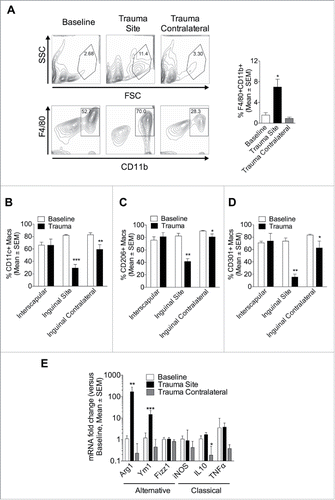
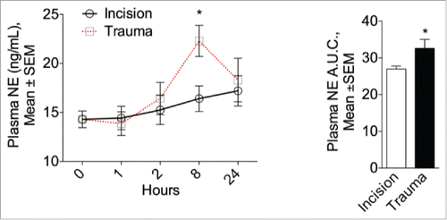
Figure 7. Surgical trauma increases plasma norepinephrine levels. Left panel: Physiological modulation of the sympathetic system in mice one, 2, 8 hours and one day after surgery or incision. NE circulating plasma levels peaked after 8 hours in the surgical trauma group but remained unaltered after incision compared to baseline (0 hour). Right panel: Quantitative assessment of plasma NE expressed as the area under the curve (A.U.C.). Data are mean±SEM of 5 independent animals per group and time point. *p < 0.05 vs. incision.