1,336
Views
8
CrossRef citations to date
0
Altmetric
Brief Report

Retinoic acid is abundantly detected in different depots of adipose tissue by SERS

, , , &
Pages 378-383 | Received 15 Aug 2016, Accepted 03 Oct 2016, Published online: 01 Nov 2016

Figures & data

Figure 1. RA pathway is differentially regulated in stem cells from VS and SC fat depots. A schematic illustrating RA synthesis / metabolism and downstream pathway regulated by RA. RA is produced in 2 sequential steps from retinol to retinal (a.k.a. retinaldehyde) by RDH10 and DHRS3, then to all-trans RA by 3 isoforms of ALDH1. WT1, a developmental marker of mesothelial layer including VS fat, controls expression of ALDH1A2 isoform. CRBP1 transports cellular retinol and retinal. Finally CYP26 family metabolizes RA into hydroxy-RA. RA acts as a major signaling molecule that binds to its receptors, RARs and RXRs. RARs then bind to its response elements (RARE), leading to modulation of various downstream target genes. Genes highlighted in red are those upregulated in VS-ASCs whereas those highlighted in blue upregulated in SC-ASCs.
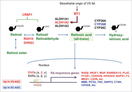
Figure 1. RA pathway is differentially regulated in stem cells from VS and SC fat depots. A schematic illustrating RA synthesis / metabolism and downstream pathway regulated by RA. RA is produced in 2 sequential steps from retinol to retinal (a.k.a. retinaldehyde) by RDH10 and DHRS3, then to all-trans RA by 3 isoforms of ALDH1. WT1, a developmental marker of mesothelial layer including VS fat, controls expression of ALDH1A2 isoform. CRBP1 transports cellular retinol and retinal. Finally CYP26 family metabolizes RA into hydroxy-RA. RA acts as a major signaling molecule that binds to its receptors, RARs and RXRs. RARs then bind to its response elements (RARE), leading to modulation of various downstream target genes. Genes highlighted in red are those upregulated in VS-ASCs whereas those highlighted in blue upregulated in SC-ASCs.

Figure 2. SERS measurement of RA levels. (A) Reaction scheme for the peroxidase reaction of TMB to TMB2+ using HRP conjugate as enzyme. Peroxidase product TMB2+ was then mixed with Au colloid for SERS measurement. (B) SERS spectra of the reactant TMB and product TMB2+ are shown for comparison. (C) RA concentration dependent SERS spectra of TMB2+. RA levels were calculated based on the intensity of 1605 cm−1 peak from TMB2+.
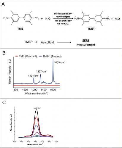
Figure 2. SERS measurement of RA levels. (A) Reaction scheme for the peroxidase reaction of TMB to TMB2+ using HRP conjugate as enzyme. Peroxidase product TMB2+ was then mixed with Au colloid for SERS measurement. (B) SERS spectra of the reactant TMB and product TMB2+ are shown for comparison. (C) RA concentration dependent SERS spectra of TMB2+. RA levels were calculated based on the intensity of 1605 cm−1 peak from TMB2+.

Figure 3. Endogenous RA levels in different tissues of mice under NC diet. Different tissues were harvested from 10-week old, male C57Bl/6J mice fed on normal chow (NC) diet (n = 4). Tissues investigated for RA concentrations by SERS were SC-WF (inguinal Subcutaneous White Fat), VS-WF (epididymal Visceral White Fat), BF (Brown Fat from the interscapular region), Eyes, Liver, SkM (quadriceps Skeletal Muscle), Heart, Sple. (Spleen), Kidn. (Kidney), Panc. (Pancreas). This result is a representative of 2 independent experiments. *p < 0.05, **p < 0.01.
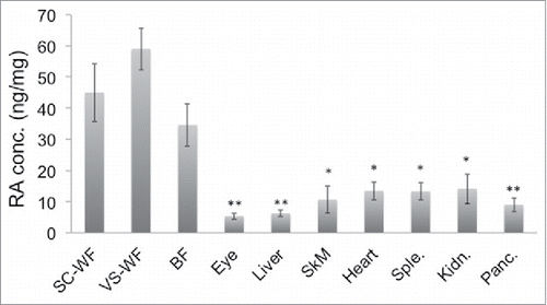
Figure 3. Endogenous RA levels in different tissues of mice under NC diet. Different tissues were harvested from 10-week old, male C57Bl/6J mice fed on normal chow (NC) diet (n = 4). Tissues investigated for RA concentrations by SERS were SC-WF (inguinal Subcutaneous White Fat), VS-WF (epididymal Visceral White Fat), BF (Brown Fat from the interscapular region), Eyes, Liver, SkM (quadriceps Skeletal Muscle), Heart, Sple. (Spleen), Kidn. (Kidney), Panc. (Pancreas). This result is a representative of 2 independent experiments. *p < 0.05, **p < 0.01.

Figure 4. Endogenous RA levels in different tissues of mice under HF diet. The same set of tissues as in were harvested from 20-week old, male C57Bl/6J mice fed on high fat (HF) diet for 12 weeks (n = 3). The tissues were then measured for their RA levels by SERS. The result shown is a representative of 2 independent experiments. *p < 0.05, **p < 0.01.
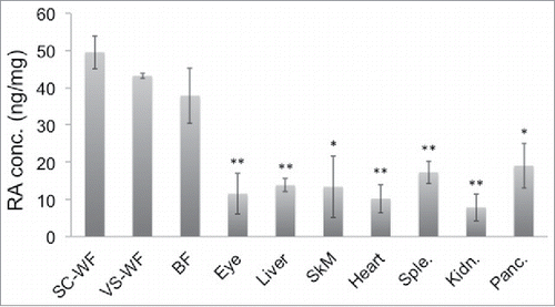
Figure 4. Endogenous RA levels in different tissues of mice under HF diet. The same set of tissues as in Figure 3 were harvested from 20-week old, male C57Bl/6J mice fed on high fat (HF) diet for 12 weeks (n = 3). The tissues were then measured for their RA levels by SERS. The result shown is a representative of 2 independent experiments. *p < 0.05, **p < 0.01.

Reprints and Corporate Permissions

Please note: Selecting permissions does not provide access to the full text of the article, please see our help page How do I view content?

To request a reprint or corporate permissions for this article, please click on the relevant link below:

Academic Permissions

Please note: Selecting permissions does not provide access to the full text of the article, please see our help page How do I view content?

Obtain permissions instantly via Rightslink by clicking on the button below:

If you are unable to obtain permissions via Rightslink, please complete and submit this Permissions form. For more information, please visit our Permissions help page.