4,621
Views
13
CrossRef citations to date
0
Altmetric
Brief Report

Lymphocytes upregulate CD36 in adipose tissue and liver

, , , , , , , , , , , , , , & show all
Pages 154-163 | Received 18 Feb 2019, Accepted 13 Apr 2019, Published online: 30 Apr 2019

Figures & data

Figure 1. CD36 expression by human adipose tissue lymphocytes. Single cells were prepared from blood, inguinal lymph node, and visceral adipose tissue stromal-vascular-fraction (AT-SVF) cells of a human donor. (a) Shown is a sample flow cytometry gating scheme of AT-SVF lymphocytes (CD3-CD4-, CD3+CD4-, and CD3+CD4+ cells, with CD41 used as a platelet discriminator in the third top dotplot from the left), and (b) HLA.DR/CD36 dotplots gated on CD3-CD4-, CD3+CD4-, and CD3+CD4+ lymphocytes of PBMC, lymph node, and AT-SVF cells, with CD36 expression based on the FMO+Isotype-FITC control. (c) Mean±sem CD36 expression by CD3-CD4-, CD3+CD4-, and CD3+CD4+ lymphocytes of human PBMC and AT-SVF cells (*p < 0.05, N = 5).
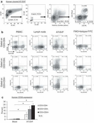
Figure 1. CD36 expression by human adipose tissue lymphocytes. Single cells were prepared from blood, inguinal lymph node, and visceral adipose tissue stromal-vascular-fraction (AT-SVF) cells of a human donor. (a) Shown is a sample flow cytometry gating scheme of AT-SVF lymphocytes (CD3-CD4-, CD3+CD4-, and CD3+CD4+ cells, with CD41 used as a platelet discriminator in the third top dotplot from the left), and (b) HLA.DR/CD36 dotplots gated on CD3-CD4-, CD3+CD4-, and CD3+CD4+ lymphocytes of PBMC, lymph node, and AT-SVF cells, with CD36 expression based on the FMO+Isotype-FITC control. (c) Mean±sem CD36 expression by CD3-CD4-, CD3+CD4-, and CD3+CD4+ lymphocytes of human PBMC and AT-SVF cells (*p < 0.05, N = 5).

Figure 2. Upregulation of CD36 expression by rhesus monkey liver and adipose tissue SVF cells. Single cells were prepared from PBMC, spleen, lymph nodes, liver, and subcutaneous and visceral AT-SVF cells of uninfected and SIV- or SHIV-infected monkeys. CD36 expression was then examined on CD3-CD8-CD4-, CD3+CD8+, and CD3+CD4+ lymphocytes by flow cytometry. Shown in A are sample CD8/CD36 and CD4/CD36 flow cytometry dotplots (gated on live CD41- lymphocytes) of an SHIV-infected monkey, and B shows mean±sem CD36 expression (*p < 0.05 compared to PBMC, spleen, or lymph nodes; lymph nodes were not examined for infected monkeys).
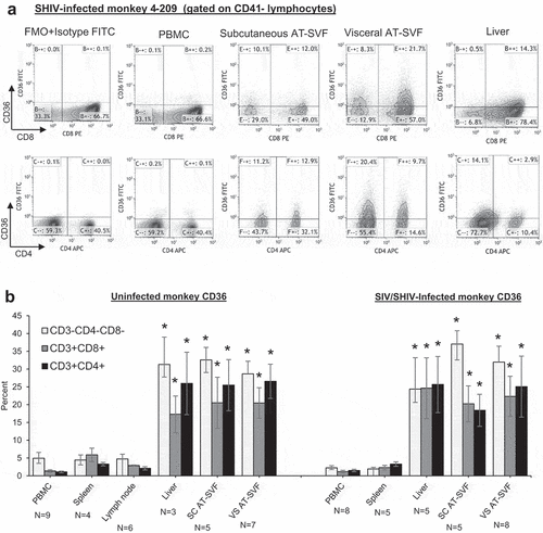
Figure 2. Upregulation of CD36 expression by rhesus monkey liver and adipose tissue SVF cells. Single cells were prepared from PBMC, spleen, lymph nodes, liver, and subcutaneous and visceral AT-SVF cells of uninfected and SIV- or SHIV-infected monkeys. CD36 expression was then examined on CD3-CD8-CD4-, CD3+CD8+, and CD3+CD4+ lymphocytes by flow cytometry. Shown in A are sample CD8/CD36 and CD4/CD36 flow cytometry dotplots (gated on live CD41- lymphocytes) of an SHIV-infected monkey, and B shows mean±sem CD36 expression (*p < 0.05 compared to PBMC, spleen, or lymph nodes; lymph nodes were not examined for infected monkeys).

Figure 3. Assessment of potential platelet interactions with T cells prepared from different tissues of monkeys. Single cells were prepared from PBMC, spleen, lymph node, liver, or subcutaneous and visceral AT-SVF cells of infected monkeys. Cells were then examined for coexpression of the CD41 platelet marker with CD3+ T cells by flow cytometry. Shown are sample CD3/CD41 dotplots of cells (gated on lymphocytes in the light scatter plot) from different tissues of four different monkeys, indicating CD3+CD41+ cells (platelet contamination of T cells) occur mostly in PBMC and spleen preparations.
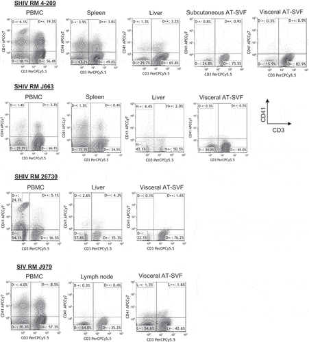
Figure 3. Assessment of potential platelet interactions with T cells prepared from different tissues of monkeys. Single cells were prepared from PBMC, spleen, lymph node, liver, or subcutaneous and visceral AT-SVF cells of infected monkeys. Cells were then examined for coexpression of the CD41 platelet marker with CD3+ T cells by flow cytometry. Shown are sample CD3/CD41 dotplots of cells (gated on lymphocytes in the light scatter plot) from different tissues of four different monkeys, indicating CD3+CD41+ cells (platelet contamination of T cells) occur mostly in PBMC and spleen preparations.

Figure 4. Predominant expression of CD36 by resting T cells in adipose tissue of rhesus monkeys. Visceral AT-SVF cells were prepared from uninfected and infected monkeys. AT-SVF CD3+ T cells were then examined for expression of CD36 in conjunction with CD38, HLA.DR, or PD-1 by flow cytometry. (a) Sample CD38/CD36, HLA.DR/CD36, and PD-1/CD36 flow cytometry dotplots (gated on CD3+CD41- T cells) of three SHIV-infected monkey AT-SVF cells. (b) Mean±sem CD36 expression by CD38-/+, HLA.DR-/+, and PD-1-/+ AT-SVF CD3+ T cells (*p < 0.05 comparing CD38-, HLA.DR-, or PD-1- cells to CD38+, HLA.DR+, or PD-1+ cells).
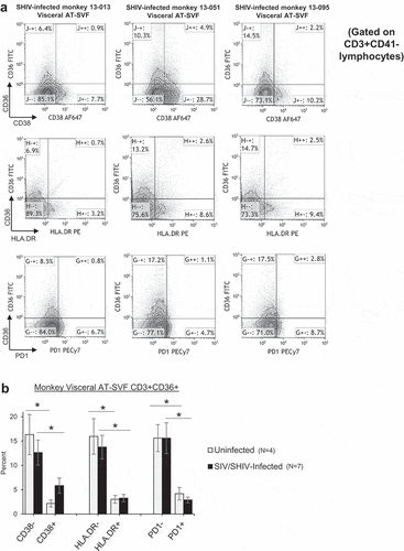
Figure 4. Predominant expression of CD36 by resting T cells in adipose tissue of rhesus monkeys. Visceral AT-SVF cells were prepared from uninfected and infected monkeys. AT-SVF CD3+ T cells were then examined for expression of CD36 in conjunction with CD38, HLA.DR, or PD-1 by flow cytometry. (a) Sample CD38/CD36, HLA.DR/CD36, and PD-1/CD36 flow cytometry dotplots (gated on CD3+CD41- T cells) of three SHIV-infected monkey AT-SVF cells. (b) Mean±sem CD36 expression by CD38-/+, HLA.DR-/+, and PD-1-/+ AT-SVF CD3+ T cells (*p < 0.05 comparing CD38-, HLA.DR-, or PD-1- cells to CD38+, HLA.DR+, or PD-1+ cells).

Figure 5. CD36 expression in adipose tissue T cells during high-fat diet in mice. (a) C57BL/6J mice were fed standard chow diet (Harlan-Teklad 2920X) or 21% milk fat/34% sucrose Breslow Western-type diet (TestDiet 5TFH) for fourteen weeks. Mice were sacrificed and single cells prepared from blood, spleen, liver, and perigonadal (PG) AT-SVF cells. Cells were then examined for CD36 expression on CD3-CD4-, CD3+CD4-, and CD3+CD4+ lymphocytes (gated on live CD41- lymphocytes) by flow cytometry. Shown are mean±sem CD36 expression (*p < 0.05). (b) Fatty acid uptake by adipose tissue CD36+ lymphocytes. PG AT-SVF cells were incubated with a fluorescent palmitate analog (C16-BODIPY), washed, and examined for C16-BODIPY uptake by CD36- and CD36+ cells (gated on CD3-CD4-, CD3+CD4-, or CD3+CD4+ lymphocytes). Shown are mean±sem C16-BODIPY mean fluorescence intensities (*p < 0.05). (c–d) Upregulation of CD36+PD-1+ lymphocytes in adipose tissue during western diet. Expression of CD36 and PD-1 by CD3-CD4-, CD3+CD4-, and CD3+CD4+ lymphocytes of spleen and PG AT-SVF cells were examined by flow cytometry. Shown are mean±sem overall PD-1 expression by spleen and PG AT-SVF lymphocytes (c), and mean±sem CD36+PD-1+ double-positive lymphocytes in PG AT-SVF cells (d, *p < 0.05).
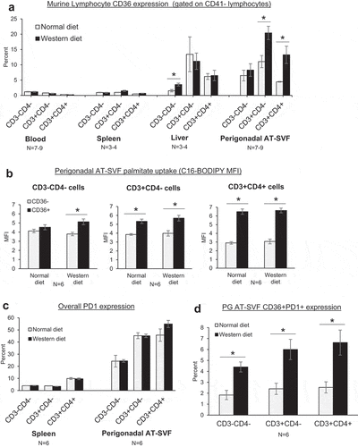
Figure 5. CD36 expression in adipose tissue T cells during high-fat diet in mice. (a) C57BL/6J mice were fed standard chow diet (Harlan-Teklad 2920X) or 21% milk fat/34% sucrose Breslow Western-type diet (TestDiet 5TFH) for fourteen weeks. Mice were sacrificed and single cells prepared from blood, spleen, liver, and perigonadal (PG) AT-SVF cells. Cells were then examined for CD36 expression on CD3-CD4-, CD3+CD4-, and CD3+CD4+ lymphocytes (gated on live CD41- lymphocytes) by flow cytometry. Shown are mean±sem CD36 expression (*p < 0.05). (b) Fatty acid uptake by adipose tissue CD36+ lymphocytes. PG AT-SVF cells were incubated with a fluorescent palmitate analog (C16-BODIPY), washed, and examined for C16-BODIPY uptake by CD36- and CD36+ cells (gated on CD3-CD4-, CD3+CD4-, or CD3+CD4+ lymphocytes). Shown are mean±sem C16-BODIPY mean fluorescence intensities (*p < 0.05). (c–d) Upregulation of CD36+PD-1+ lymphocytes in adipose tissue during western diet. Expression of CD36 and PD-1 by CD3-CD4-, CD3+CD4-, and CD3+CD4+ lymphocytes of spleen and PG AT-SVF cells were examined by flow cytometry. Shown are mean±sem overall PD-1 expression by spleen and PG AT-SVF lymphocytes (c), and mean±sem CD36+PD-1+ double-positive lymphocytes in PG AT-SVF cells (d, *p < 0.05).
Supplemental material