Figures & data

Figure 1. Effects of LIPUS on rat preadipocyte differentiation and proliferation. Primary-cultured preadipocytes were treated with different doses of ultrasound (0.8, 1.0 and 1.2 MPa) for 10 min. (a) Preadipocyte differentiation after different dose of LIPUS treatment was visualized by the oil red O staining. (b) Quantification of lipid droplet formation after different dose of LIPUS treatment. (c,d) Cell proliferation ability was analyzed by flow cytometric analysis using cell-light EdU Apollo 488 kit staining. All values are expressed as the mean ± SEM of three independent trials. Data were analyzed with independent t test. *** p < 0.001. Bar: 50 μm.
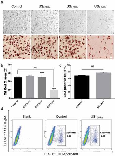
Figure 2. Effects of LIPUS on gene expression associated with adipogenic differentiation. Cells were prepared for qPCR and western after 2 d of adding adipogenic medium. We chose this time point because the levels of C/EBPs and PPARγ will be obviously up-regulated in the stage of initial differentiation and the levels of C/EBP beta and delta will not change in the stage of terminal differentiation compared to the stage of preadipocyte. (a) The mRNA levels of PPARγ, C/EBPα, C/EBPβ and C/EBPδ were examined by qRT-PCR assay in differentiated preadipocytes after LIPUS treatment. (b,c) The protein expression levels of PPARγ and C/EBPα were assessed by western blotting in differentiated preadipocytes after LIPUS treatment. (d) The mRNA levels of representative adipogenic markers including Fabp4 and adiponectin after LIPUS treatment were examined by qRT-PCR. (e) Preadipocytes were treated with the PPARγ agonist ROZ (1 μM). The effects of ROZ on the LIPUS-induced inhibition of preadipocyte differentiation were measured by oil red O staining. (f) The effects of ROZ on adipogenic differentiation markers after LIPUS treatment were examined by qRT-PCR. All values are expressed as the mean ± SEM of three independent trials. Data were analyzed with independent t test. * p< 0.05, ** p< 0.01, *** p< 0.001. Bar: 50 μm.
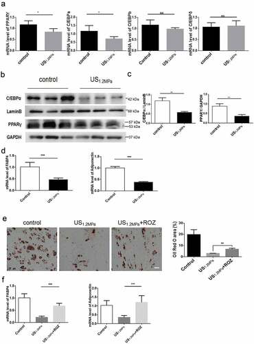
Figure 3. LIPUS increases HDAC1 expression and decreases histone 3 and histone 4 acetylation modification. (a) The phosphorylation levels of ERK, P38 and JNK were analyzed by western blotting. (b) Quantification of p-ERK, p-P38 and p-JNK normalized to their total proteins. (c) Nuclear protein levels of HDAC1, HDAC2, HDAC6, AcH3 and AcH4 were assessed by western blotting. (d) Quantification of HDAC1, HDAC2, HDAC6, AcH3 and AcH4 normalized to Lamin B. Relative protein levels were analyzed with independent t test. All values are expressed as the mean ± SEM of three independent trials. * P < 0.05.
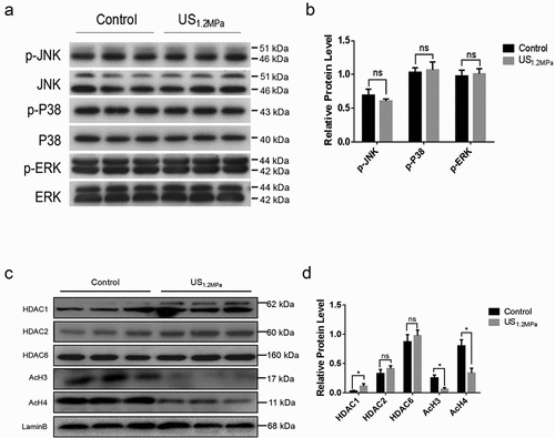
Figure 4. SAHA could rescue the effects of LIPUS on adipogenic differentiation. Preadipocytes were treated with LIPUS combined with the HDACs inhibitor SAHA. (a) The effects of HDAC1 inhibition on preadipocyte differentiation after LIPUS treatment were evaluated by the oil red O staining. (b) The effects of HDAC1 inhibition on protein levels of PPARγ, C/EBPα, HDAC1, AcH3 and AcH4 after LIPUS treatment were assessed by western blotting. Quantification of indicated proteins normalized to lamin B(HDAC1, C/EBPα, AcH3 and AcH4) or GAPDH(PPARγ). (c) The effects of HDAC1 inhibition on the mRNA expression of adipogenic differentiation markers after LIPUS treatment were examined by qRT-PCR assay. (d) The effect of GW9662 on the SAHA’s effect after LIPUS treatment were assessed by the oil red O staining. All values are expressed as the mean ± SEM of three independent trials. Data were analyzed with independent t test. * p< 0.05, ** p< 0.01, *** p< 0.001, **** p< 0.0001. Bar: 50 μm.
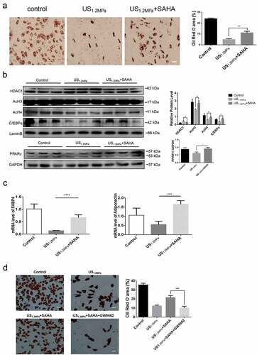
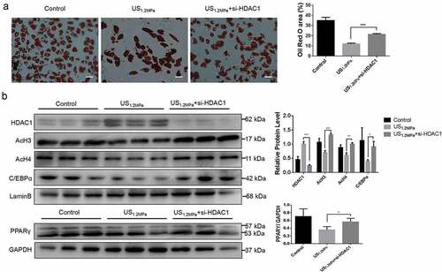
Figure 5. Down-regulation of HDAC1 by siRNA reverses the effects of LIPUS on adipogenic differentiation. Preadipocytes were treated with LIPUS combined with the si-HDAC1. (a) The effects of HDAC1 inhibition by si-RNA on preadipocyte differentiation after LIPUS treatment were evaluated by the oil red O staining. (b) The effects of HDAC1 inhibition by si-RNA on protein levels of PPARγ, C/EBPα, HDAC1, AcH3 and AcH4 after LIPUS treatment were assessed by western blotting. Quantification of indicated proteins normalized to lamin B(HDAC1, C/EBPα, AcH3 and AcH4) or GAPDH(PPARγ). All values are expressed as the mean ± SEM of three independent trials. Data were analyzed with independent t test. * p< 0.05, ** p< 0.01, *** p< 0.001. Bar: 50 μm.