
Figures & data
Figure 1. Genetic transduction of adipose-derived stem cells. (a) Representative photomicrograph of adipose-derived stem cells transducted with adenovirus encoding GFP gene. (b) Representative Western blots showing overexpression of Bcl-2 protein in Bcl-2 modified adipose-derived stem cells, which remained at a high level for 21 d after transduction. Housekeeping protein β-actin served as loading control. Scale bars = 100 μm.
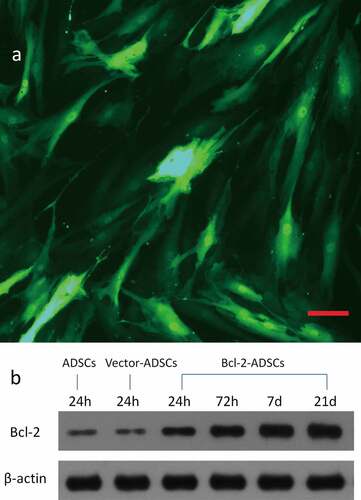
Figure 2. Retention of the fat grafts 6 weeks after transplantation. (a) Surviving fat grafts under the skin of a representative nude mouse. (b) Harvested fat grafts. (c) Mass of fat grafts was higher in the Fat + Bcl-2-modified adipose-derived stem cells group (Fat + Bcl-2-ADSCs) than in the Fat-only group, or than in the Fat + vector-modified adipose-derived stem cells group (Fat + Vector-ADSCs). The differences between any two groups were statistically significant.
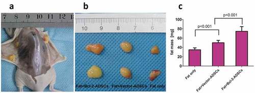
Figure 3. Histological features of transplanted fat tissues. Grafts from Fat + Bcl-2-modified adipose-derived stem cells group (Fat + Bcl-2-ADSCs) contained more adipocytes and lower levels of fat necrosis and fibrosis than those from the Fat-only group, or than those from Fat + vector-modified adipose-derived stem cells group (Fat + Vector-ADSCs). Scale bars = 50 µm.
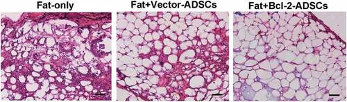
Figure 4. Bcl-2-modified adipose-derived stem cells in transplanted fats. (a) Representative images from immunostaining of transplanted adipose-derived stem cells in the Fat + vector-modified adipose-derived stem cells group (Fat + Vector-ADSCs) and in the Fat + Bcl-2-modified adipose-derived stem cells group (Fat + Bcl-2-ADSCs) 3 weeks and 6 weeks after transplantation. BrdU-labelled adipose-derived stem cells (brown) were clearly identified. (BrdU) (brown). (DAPI) (blue). Representative cell engraftment was detected (white arrows). (b) Quantitative assessment of surviving adipose-derived stem cells at 3 weeks, and 6 weeks. The number of surviving Bcl-2-modified adipose-derived stem cells was greater than vector-modified adipose-derived stem cells at each time point. Scale bars = 20 µm.
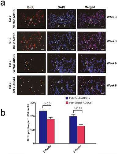
Figure 5. The angiogenic differentiation of Bcl-2-modified adipose-derived stem cells 3 weeks after transplantation. The vWF-positive vascular structure was red. The BrdU-positive Bcl-2-modified adipose-derived stem cells were green. In merged image of the vWF-positive vascular structure with the BrdU-positive Bcl-2-modified adipose-derived stem cells, arrows indicate the location of BrdU-positive and vWF-positive cells. Double immunofluorescence staining revealed Bcl-2-modified adipose-derived stem cells incorporated into the endothelial lining of neocapillary. Scale bars = 20 µm.
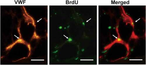
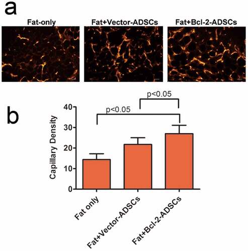
Figure 6. Bcl-2-modified adipose-derived stem cells enhanced angiogenesis in the transplanted fats. (a) Representative photomicrographs of the transplanted fats obtained after immunostaining for vWF antibody. (b) Quantitative capillary density data based on vWF immunostaining. Capillary density was much higher in the Fat + Bcl-2-modified adipose-derived stem cells group (Fat + Bcl-2-ADSCs) than in the Fat-only group, or Fat + vector-modified adipose-derived stem cells group (Fat + Vector-ADSCs). The differences between any two groups were statistically significant. Scale bars = 20 µm.
Data availability statement
The datasets used and/or analysed during the present study are available from the corresponding author on reasonable request (https://doi.org/10.1080/21623945.2022.2107195).