Figures & data
FIGURE 1. IL-6 and downstream signaling molecule expression in hearts of Ryr2+/+ and Ryr2ADA/ADA mice. (A) Immunoblots of heart homogenates from 1-day old and 10-day old Ryr2+/+ (WT) and Ryr2ADA/ADA (ADA) mice. Glyceraldehyde-3-phosphate dehydrogenase (GAPDH) was the loading control. (B) Relative protein and phosphorylation levels in 1-day old and 10-day old Ryr2ADA/ADA compared to Ryr2+/+ mice. Data are the mean ± SEM of 5–19 samples. *p < 0.05 compared to Ryr2+/+, #p < 0.05 compared to corresponding 1 day sample using one way ANOVA.
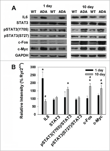
FIGURE 2. Nuclear and cytosolic location of STAT3 and pSTAT3-Tyr705. (A) Immunoblots of nuclear (N) and cytosolic (C) fractions from hearts of 10-day old Ryr2+/+ (WT) and Ryr2ADA/ADA (ADA) mice. GAPDH and histone 3 (His3) were markers for cytosolic and nuclear fractions, respectively. (B) Protein and phosphorylation levels of Ryr2ADA/ADA mice were normalized to Ryr2+/+. Data are the mean ± SEM of 5–6 samples.*p < 0.05 compared to Ryr2+/+ using t-test.
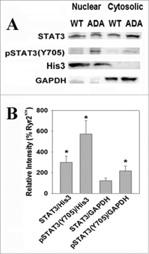
FIGURE 3. Ryr2ADA/ADA and Ryr2ADA/ADA/IL-6−/− mice survival. Mean lifetimes ± SEM of Ryr2ADA/ADA and Ryr2ADA/ADA/IL-6−/− mice of 16.9 ± 1.3 (n = 9) and 25.1 ± 3.5 (n = 7), respectively, were significantly different (p < 0.05).
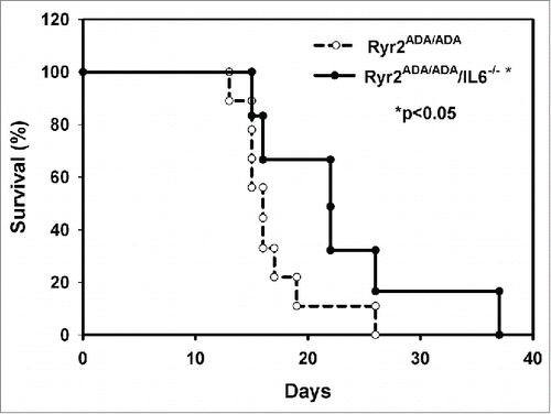
Table 1. Body and heart weights and echocardiography of 10-day old Ryr2+/+ and Ryr2ADA/ADA mice.
FIGURE 4. Expression of IL-6 downstream signaling molecules in mice targeted for Ryr2ADA and IL-6−/−. (A) Immunoblots of heart homogenates from 10-day old Ryr2+/+ (WT) and Ryr2ADA/ADA (ADA) mice with and without IL-6. GAPDH was the loading control. (B) Protein levels and phosphorylation ratios of Ryr2ADA/ADA mice were normalized to Ryr2+/+. Data are the mean ± SEM of 6–14 determinations. Footnotep < 0.05 compared with Ryr2+/+, #p < 0.05 compared with Ryr2ADA/ADA, using one way ANOVA.
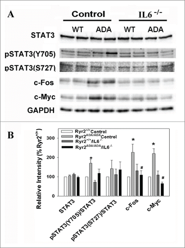
FIGURE 5. Survival data from Ryr2ADA/ADA mice treated with STAT3 inhibitor NSC74859. Mean lifetimes ± SEM of Ryr2ADA/ADA mice treated without (Control) and with NSC74859 (NSC) of13.9 ± 0.5 (n = 13) and 18.4 ± 1.8 (n = 13), respectively, were significantly different (p < 0.05).
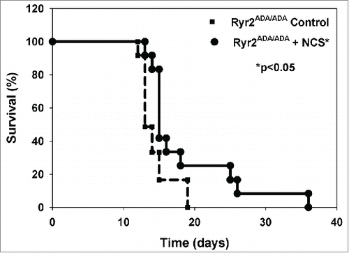
Table 2. Body and heart weights and echocardiography of 10-day old Ryr2+/+ and Ryr2ADA/ADA mice with and without NSC74859.
FIGURE 6. IL-6 and downstream signaling molecule levels in 10-day old mice treated with or without STAT3 inhibitor NSC74859. (A) Immunoblots of heart homogenates from 10-day old Ryr2+/+ (WT) and Ryr2ADA/ADA (ADA) mice treated with or without NSC74859 (NSC). GAPDH was the loading control. (B) Protein levels and phosphorylation ratios of Ryr2ADA/ADA mice were normalized to Ryr2+/+ Control. Data are the mean ± SEM of 4–10 samples. *p < 0.05 compared to Ryr2+/+ mice without NSC74859, #p < 0.05 compared to Ryr2ADA/ADA mice without NSC74859, using one way ANOVA.
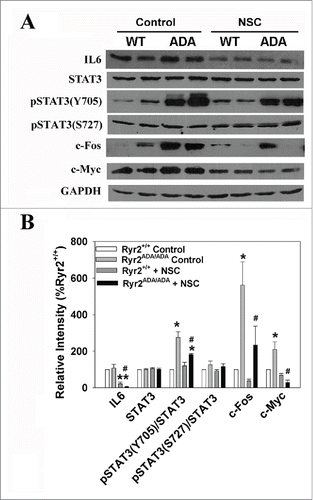
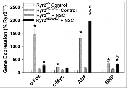
FIGURE 7. c-Fos, c-Myc, ANP and BNP mRNA levels in hearts of Ryr2+/+ and Ryr2ADA/ADA mice treated with or without STAT3 inhibitor NSC74859. mRNA levels were measured by quantitative RT-PCR and normalized to levels in hearts of Ryr2+/+ mice not treated with the inhibitor (Ryr2+/+ Control). Data are the mean ± SEM of 5–6 samples. *p < 0.05 compared to Ryr2+/+ mice without NSC74859, #p < 0.05 compared with Ryr2ADA/ADA mice without NSC74859, %p < 0.05 compared with Ryr2+/+ mice with NSC74859, using one way ANOVA.