1,643
Views
53
CrossRef citations to date
0
Altmetric
Original Research

Enhanced neutrophil activity is associated with shorter time to tumor progression in glioblastoma patients

, , , , , , , , , , & show all
Article: e1075693 | Received 20 Apr 2015, Accepted 17 Jul 2015, Published online: 26 Feb 2016

Figures & data

Figure 1. Neutrophil activity was enhanced in 12 patients and was unchanged or decreased in 16 patients over time (Group I;GI with enhanced neutrophil activity: T0 vs. T24W: p < 0.0001 and T12W vs. T24W: < 0.0001, Group II; GII with unchanged or decreased neutrophil activity: T12W vs. T24W: = 0.014, ). Kaplan–Meier survival curves of GBM patients and grade of HCMV infection in their tumors. (B and C) GBM patients with high neutrophil activity (GI) had significantly shorter TTP (A) and shorter median OS (B) than patients with unchanged or decreased neutrophil activity (GII). (D) Significantly more patients in GI had high-grade HCMV infection (Grades 3+, 4+) in their tumor compared with GII (p = 0.047).
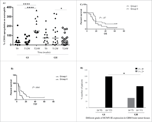
Figure 1. Neutrophil activity was enhanced in 12 patients and was unchanged or decreased in 16 patients over time (Group I;GI with enhanced neutrophil activity: T0 vs. T24W: p < 0.0001 and T12W vs. T24W: p < 0.0001, Group II; GII with unchanged or decreased neutrophil activity: T12W vs. T24W: p = 0.014, Fig. 1A). Kaplan–Meier survival curves of GBM patients and grade of HCMV infection in their tumors. (B and C) GBM patients with high neutrophil activity (GI) had significantly shorter TTP (A) and shorter median OS (B) than patients with unchanged or decreased neutrophil activity (GII). (D) Significantly more patients in GI had high-grade HCMV infection (Grades 3+, 4+) in their tumor compared with GII (p = 0.047).

Figure 2. Levels of 12 cytokines, chemokines, and inflammatory factors in plasma samples from GBM patients in Group I (GI) and Group II (GII) and healthy controls (HC) (A–F).The levels of IL-12p70 was significantly increased over time (T0 vs. T24W, p = 0.002) only in Group I patients with significantly enhanced neutrophil activation (A). The levels of IL-8 and MCP-1 were significantly elevated overtime in both Group I and Group II compared with healthy controls (HC) (IL-8: GI: T0 vs. HC, p = 0.0009; T12W vs. HC, p = 0.008 and T24W vs. HC, p = 0.0009 and GII: T0 vs. HC, p <0.0001; T12W vs. HC, p = 0.0002 and T24W vs. HC, p = 0.0007) (MCP-1: GI: T0 vs. HC, p = 0.0002; T12W vs. HC, p = 0.0002; T24W vs. HC, p = 0.0002 and GII: To vs. HC, = 0.0002; T12W vs. HC, = 0.0002 and T24W vs. HC, = 0.0005) (B, C). However the level of IL-8 was deceased at 12 weeks (p = 0.02) (D) only in Group II with decreased or unchanged neutrophil activity. The levels of MCP-1 and IL-8 did not differ between GI and GII (B–C). Other examined factors did not differ between the two groups (A–F).
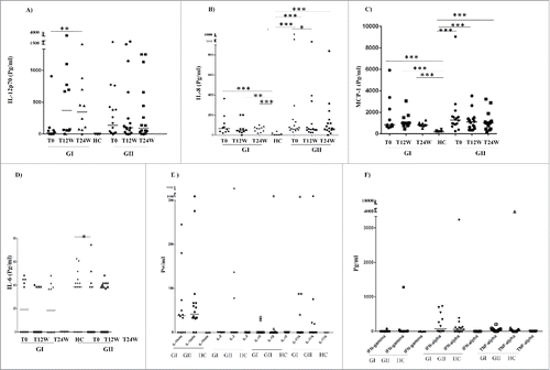
Figure 2. Levels of 12 cytokines, chemokines, and inflammatory factors in plasma samples from GBM patients in Group I (GI) and Group II (GII) and healthy controls (HC) (A–F).The levels of IL-12p70 was significantly increased over time (T0 vs. T24W, p = 0.002) only in Group I patients with significantly enhanced neutrophil activation (A). The levels of IL-8 and MCP-1 were significantly elevated overtime in both Group I and Group II compared with healthy controls (HC) (IL-8: GI: T0 vs. HC, p = 0.0009; T12W vs. HC, p = 0.008 and T24W vs. HC, p = 0.0009 and GII: T0 vs. HC, p <0.0001; T12W vs. HC, p = 0.0002 and T24W vs. HC, p = 0.0007) (MCP-1: GI: T0 vs. HC, p = 0.0002; T12W vs. HC, p = 0.0002; T24W vs. HC, p = 0.0002 and GII: To vs. HC, p = 0.0002; T12W vs. HC, p = 0.0002 and T24W vs. HC, p = 0.0005) (B, C). However the level of IL-8 was deceased at 12 weeks (p = 0.02) (D) only in Group II with decreased or unchanged neutrophil activity. The levels of MCP-1 and IL-8 did not differ between GI and GII (B–C). Other examined factors did not differ between the two groups (A–F).

Figure 3. Median levels of 12 cytokines/chemokines, and inflammatory factors in plasma of all GBM patients over time and in healthy controls (HC) (3A–F). The median levels of IL-1β, IL-6, IL-8, IL-12p70, IFN-α TNF-α, MCP-1, and TGF-β (but not IFNγ, IL-2, IL-10, and IL-17A) were higher at baseline in GBM patients than in healthy controls (). During the 24 weeks of antitumor treatment, GBM patients had significantly decreased levels of IL-6 (T0 vs. T12, p = 0.04) (B) and increased levels of IL-12p70 (T0 vs. T12, p = 0.04) (C), significantly decreased levels of MCP-1 (T0 vs. T12, p = 0.008; T0 vs. T24, p = 0.03) (E) and TGF-β (T0 vs. T12, p = 0.0006 and T0 vs. T24, p < 0.0001, T12W vs. T24W, p = 0.04) (F).
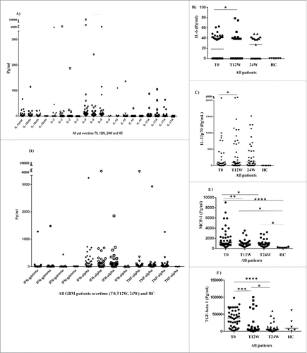
Figure 3. Median levels of 12 cytokines/chemokines, and inflammatory factors in plasma of all GBM patients over time and in healthy controls (HC) (3A–F). The median levels of IL-1β, IL-6, IL-8, IL-12p70, IFN-α TNF-α, MCP-1, and TGF-β (but not IFNγ, IL-2, IL-10, and IL-17A) were higher at baseline in GBM patients than in healthy controls (Fig. 3A–F). During the 24 weeks of antitumor treatment, GBM patients had significantly decreased levels of IL-6 (T0 vs. T12, p = 0.04) (B) and increased levels of IL-12p70 (T0 vs. T12, p = 0.04) (C), significantly decreased levels of MCP-1 (T0 vs. T12, p = 0.008; T0 vs. T24, p = 0.03) (E) and TGF-β (T0 vs. T12, p = 0.0006 and T0 vs. T24, p < 0.0001, T12W vs. T24W, p = 0.04) (F).

Figure 4. Median levels of 12 examined pro-inflammatory cytokines and chemokines in the plasma of GBM patients treated with valganciclovir (V) or placebo (P) over time (A–I). The cytokine profiles in these two groups did not differ (), except for IL-12p70 and IL-8. IL-12p70 levels decreased significantly in valganciclovir-treated patients (T12 vs. T24, p = 0.03 (E) but increased significantly in placebo patients (T0 vs. T24, p = 0.004) (C). The level of IL-8 decreased only in the placebo group at 12 weeks (T0 vs. T12, p = 0.008) (B).
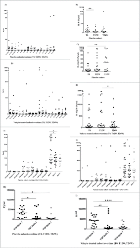
Figure 4. Median levels of 12 examined pro-inflammatory cytokines and chemokines in the plasma of GBM patients treated with valganciclovir (V) or placebo (P) over time (A–I). The cytokine profiles in these two groups did not differ (Fig. 4A, D, F, G, H, I), except for IL-12p70 and IL-8. IL-12p70 levels decreased significantly in valganciclovir-treated patients (T12 vs. T24, p = 0.03 (E) but increased significantly in placebo patients (T0 vs. T24, p = 0.004) (C). The level of IL-8 decreased only in the placebo group at 12 weeks (T0 vs. T12, p = 0.008) (B).
Supplemental material

Supplemental_Material.zip

Download Zip (898.3 KB)

Reprints and Corporate Permissions

Please note: Selecting permissions does not provide access to the full text of the article, please see our help page How do I view content?

To request a reprint or corporate permissions for this article, please click on the relevant link below:

Academic Permissions

Please note: Selecting permissions does not provide access to the full text of the article, please see our help page How do I view content?

Obtain permissions instantly via Rightslink by clicking on the button below:

If you are unable to obtain permissions via Rightslink, please complete and submit this Permissions form. For more information, please visit our Permissions help page.