Figures & data
Figure 1. Th22 cell-derived IL-22 stimulates colon cancer cell proliferation. (A) GSEA analysis in the association between IL-22 and cell proliferation pathways in the TCGA colon cancer dataset. n = 224, nominal p < 0.05, false discovery rate [FDR] q < 0.25, red bar: positively correlated genes, blue bar: negatively correlated genes. (B) Effect of endogenous IL-22 on primary colon cancer cell proliferation. Single cells including colon cancer cells and immune cells were isolated from fresh colon cancer tissue and cultured with or without anti-IL-22 antibody for 24 h. Cell proliferation was tested by H3 Thymidine Incorporation. Results are expressed as the mean of CPM ± SD. One of three patients with triplicates is shown. *p < 0.05. (C) Effect of endogenous Th-22-derived IL-22 on the established primary colon cancer cell proliferation. Freshly sorted colon cancer associated CD4+ T cells were stimulated with anti-CD3 and anti-CD28 for 3 d. Established primary colon cancer cells (C1) were cultured with these T cell supernatants with or without anti-IL-22 antibody for 24 h. Tumor cell proliferation was evaluated with thymidine incorporation. Results are expressed as the mean of CPM ± SD. One of three patients with triplicates is shown. *p <0.05. (D–F) Effect of exogenous IL-22 on colon cancer cell proliferation. DLD-1 (D-F), HT29 (F), C1 and C2 primary colon cancer cells (F) were treated with or without recombinant IL-22 for 24 h. Cell proliferation was evaluated with Ki67 expression by FACS (D) and thymidine incorporation (E). The absolute cell numbers were counted and relative cell numbers were shown (F). One of four repeats is shown. *p <0.05.
![Figure 1. Th22 cell-derived IL-22 stimulates colon cancer cell proliferation. (A) GSEA analysis in the association between IL-22 and cell proliferation pathways in the TCGA colon cancer dataset. n = 224, nominal p < 0.05, false discovery rate [FDR] q < 0.25, red bar: positively correlated genes, blue bar: negatively correlated genes. (B) Effect of endogenous IL-22 on primary colon cancer cell proliferation. Single cells including colon cancer cells and immune cells were isolated from fresh colon cancer tissue and cultured with or without anti-IL-22 antibody for 24 h. Cell proliferation was tested by H3 Thymidine Incorporation. Results are expressed as the mean of CPM ± SD. One of three patients with triplicates is shown. *p < 0.05. (C) Effect of endogenous Th-22-derived IL-22 on the established primary colon cancer cell proliferation. Freshly sorted colon cancer associated CD4+ T cells were stimulated with anti-CD3 and anti-CD28 for 3 d. Established primary colon cancer cells (C1) were cultured with these T cell supernatants with or without anti-IL-22 antibody for 24 h. Tumor cell proliferation was evaluated with thymidine incorporation. Results are expressed as the mean of CPM ± SD. One of three patients with triplicates is shown. *p <0.05. (D–F) Effect of exogenous IL-22 on colon cancer cell proliferation. DLD-1 (D-F), HT29 (F), C1 and C2 primary colon cancer cells (F) were treated with or without recombinant IL-22 for 24 h. Cell proliferation was evaluated with Ki67 expression by FACS (D) and thymidine incorporation (E). The absolute cell numbers were counted and relative cell numbers were shown (F). One of four repeats is shown. *p <0.05.](/cms/asset/e250fec0-b193-4fed-bb49-42041edcd2b9/koni_a_1082704_f0001_oc.gif)
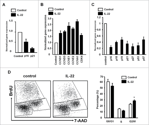
Figure 2. IL-22 targets cyclin genes and controls colon cancer cell proliferation. (A–C) Effect of IL-22 on the expression of cell proliferation genes in colon cancer cells. Colon cancer cells were cultured with IL-22 for 12 h. The levels of cyclin-dependent kinase inhibitors and cyclins were quantified by real-time PCR in DLD-1 (A, B) and primary colon cancer cells (C). Results are expressed as the relative values (mean ± SD). One of three experiments with triplicates is shown. p < 0.05. (D) Effect of IL-22 on colon cancer cell cycles. DLD-1 cells were cultured with IL-22 for 24 h. Expression of BrdU and 7-AAD was analyzed by FACS. Results are shown as the percent of cells in G0/G1, S, G2/M. n = 4, p <0.05.

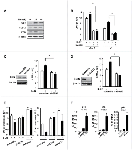
Figure 3. IL-22 induced colon cancer proliferation depends on H3K27me3, not H3K79me2. (A) Effect of IL-22 on PCR2 component expression in colon cancer cells. DLD-1 cells were cultured with IL-22 (20ng/mL) for 24 or 48 h. Expression of EZH2, EED, SUZ12 was detected by Western blotting. One of three experiments is shown. (B) Effect of DZNep on IL-22-induced colon cancer cell proliferation. DLD-1 and C1, primary colon cancer cells were treated with IL-22 and EZH2 inhibitor DZNep for 24 h. Cell proliferation was detected by H3 thymidine incorporation. n = 4, p < 0.05. (C, D) Effect of shEZH2 and shSUZ12 on DLD-1 cell proliferation. DLD-1 cells were transfected with control vector (scramble), shEZH2 (C) and shSUZ12 (D), and stimulated with IL-22 for 24 h. EZH2 expression was detected by Western blotting (left). Cell proliferation was detected by H3 thymidine incorporation (right). n = 4, * p < 0.05. (E) Effect of shEZH2 and shSUZ12 on the expression of p16 and p21 in DLD-1 cells. DLD-1 cells were transfected with control vector (scramble), shEZH2 and shSUZ12, and stimulated with IL-22 for 24 h. P16 and p21 expression was quantified by real-time PCR. n = 3, p < 0.05. (F) Effect of IL-22 on the occupancies of H3K27me3 in the promoters of p16 and p21 in DLD-1 cells. DLD-1 cells were cultured with IL-22 for 24 h. The occupancies of H3K27me3 in the promoters of p16 and p21 were determined by ChIP assays. n = 3, * p < 0.05.

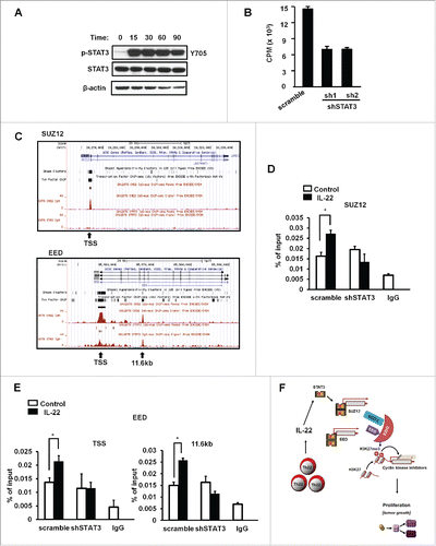
Figure 4. IL-22 promoted STAT3 binding to the PRC2 promoters. (A) Activation of STAT3 by IL-22. Colon cancer cells were treated with IL-22 for different time points. Phosphorylated STAT3 and STAT3 proteins were detected by immunoblotting. One of three experiments is shown. (B) Effect of shSTAT3 on IL-22-induced DLD-1 cell proliferation. DLD-1 cells were transfected with control vector (scramble) or two vectors encoding shSTAT3, and stimulated with IL-22 for 24 h. Cell proliferation was detected by H3 thymidine incorporation. n = 3, p < 0.05. (C) Based on the ENCODE STAT3-ChIP-Seq data base, STAT3 occupancy on the promoter areas of SUZ12 and EED is shown. (D, E) Effect of IL-22 on the occupancy of STAT3 in the promoters of PRC2 components in DLD-1 cells. DLD-1 cells expressing control vector or shSTAT3 were cultured with IL-22 for 0.5 h. The occupancy of STAT3 in the promoters of SUZ12 (D) and EED (E) was determined by ChIP assays. n = 3, p < 0.05. (F) Scheme of the mode of action of Th22 cells in colon cancer cell proliferation. Th22 cells release IL-22, activate STAT3, and STAT3 binds to the promoters of PRC2 components causing tumor cell proliferation via H3K27me3-mediated p16 and p21 repression.

