Figures & data
Figure 1. ALA-induced tumor growth inhibition depends on both cancer and host TLR4. (A) Tumor growth curve of FvB mice bearing subcutaneous NT2 cells after treatment or not with ALA (8 mg/kg) (8/group). Statistical test: Mann–Whitney U-test. **p < 0.01, ***p < 0.001. (B–C) mice survival curves of (B) Balb/c mice bearing EMT-6H cells, in wild-type (wt) mice or mice with the TLR4 gene knocked-out (TLR4−/−) after treatment or not with ALA (8 mg/kg) (20/group) or (C) mice bearing EMT-6H cells knocked-down for TLR4 (clone A and B) after treatment or not with ALA (8 mg/kg) (10/group). Survival curves were statistically different (***p <0.001 Log–Rank (Mantel–Cox) test). (D) Quantification of ALA, as measured by β-hydroxymyristic acid analysis, in different mice tissues 24 h after the 3rd ALA injection. Organs and lung nodules were taken from 27 treated and 12 control mice. Data are representative of at least two experiments. Student's t test *p < 0.05, **p < 0.01, ***p < 0.001, ****p < 0.0001.
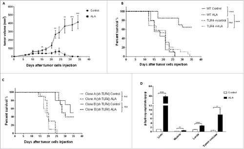
Figure 2. ALA-induced tumor growth inhibition depends on both IFNγ and NOS II. (A) Wild-type (wt) Balb/c mice or mice with the IFNγ gene knocked-out (IFNγ−/−) bearing EMT-6H tumors were treated with ALA (8 mg/kg) or not (control) and mice survival was monitored for 35 d (10/group) (***p < 0.001, Log–Rank (Mantel–Cox) test). (B) Clones A and B of shIFNR2 EMT-6H cells were injected into wild-type mice before treatment or not with ALA (8 mg/kg). Mice survival was monitored for 35 d (10/group). Survival curves were statistically non-significant (p > 0.05 Log–Rank (Mantel–Cox) test). Data are representative of two experiments. (C) Balb/c mice with the NOS II gene knocked-out (NOS II−/−) and bearing EMT-6H tumors were treated as in (B), in the presence or absence of the NOS II inhibitor aminoguanidine (AG, 50 mg/kg). Mice survival was monitored for 35 d (20/group).*p < 0.05 Log–Rank (Mantel–Cox) test)). (D) Western blot analysis of the protein expression of NOS isoforms in EMT-6H cells treated or not with ALA (500 ng/mL) and IFNγ (33 ng/mL). Brain lysate and human umbilical vein endothelial cells (HUVEC) were used as positive controls. HSC70 served as a loading control.
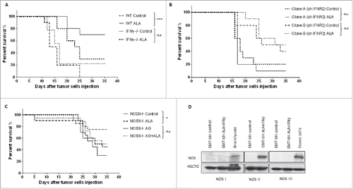
Figure 3. ALA and IFNγ induce cell death in EMT-6H cells in vitro through TLR4, IFNR and NOS II. (A) Cell viability analysis of clones A and B of TLR4 deficient EMT-6H cells or not after 48 h of cell incubation with ALA (500 ng/mL) and IFNγ (33 ng/mL). (B) Cell viability analysis of clones A and B of EMT-6H cells deleted or not for IFNR2 after 48 h treatment with ALA plus IFNγ as in (A). (C) Effects of NOS II inhibitors (aminoguanidine [AG; 0.5 mM] and 1400 W; 10 µM) on cell viability induced by ALA plus IFNγ. Data (A–C) are the mean of at least three independent experiments. Student's t test *p < 0.05. (D–F) Western blot analysis of NOS II expression induced by ALA and/or IFNγ as in (A) in WT EMT-6H cells (D) or those deleted for TLR4 (sh TLR4) or INFR2 (sh IFNR2) (E). Data are representative of three independent experiments. HSC70 served as a loading control.
![Figure 3. ALA and IFNγ induce cell death in EMT-6H cells in vitro through TLR4, IFNR and NOS II. (A) Cell viability analysis of clones A and B of TLR4 deficient EMT-6H cells or not after 48 h of cell incubation with ALA (500 ng/mL) and IFNγ (33 ng/mL). (B) Cell viability analysis of clones A and B of EMT-6H cells deleted or not for IFNR2 after 48 h treatment with ALA plus IFNγ as in (A). (C) Effects of NOS II inhibitors (aminoguanidine [AG; 0.5 mM] and 1400 W; 10 µM) on cell viability induced by ALA plus IFNγ. Data (A–C) are the mean of at least three independent experiments. Student's t test *p < 0.05. (D–F) Western blot analysis of NOS II expression induced by ALA and/or IFNγ as in (A) in WT EMT-6H cells (D) or those deleted for TLR4 (sh TLR4) or INFR2 (sh IFNR2) (E). Data are representative of three independent experiments. HSC70 served as a loading control.](/cms/asset/75cac8dc-1e49-4770-9579-8c784c237b4e/koni_a_1123369_f0003_b.gif)
Figure 4. ALA plus IFNγ induce NO and ROS production necessary but not sufficient by themselves to induce tumor cell toxicity. (A) Analysis of EMT-6H cell death after treatment with ALA (500 ng/mL), IFNγ(33 ng/mL) or cPTIO(2-(4-carboxyphenyl)-4,4,5,5-tetramethylimidazoline-1-oxyl-3-oxide; 50 µM) for 48 h. The data of one experiment representative of five independent ones are shown. Student's t test ****p < 0.0001. (B) Cell death analysis (in black) of EMT-6H cells and NO production (in white) as attested by nitrite accumulation after treatment for 48 h with different concentrations of GTN (Glyceryltrinitrate). (C) EMT-6H cells were treated with ALA (500 ng/mL) and IFNγ (33 ng/mL) in the presence or absence of 10 µM 1400 W for 24 h. ROS production was detected by flow cytometry after adding the DCFH-2-DA probe for 30 min. (D) EMT-6H cells were treated as in (C). Production of superoxide was detected by Electronic paramagnetic resonance after adding CMH for 15 min. Data show the mean light intensity emitted and are representative of two independent experiments. (E) Mice were treated as described in . Lung tissues were harvested after the 3rd ALA injection and cryo-sections were incubated with DHE (dihydroethidium), which fluoresces red in the presence of ROS. Cell nuclei were stained with DAPI (blue). Scale bars: 20 μm. (F) Survival curve of Balb/c mice bearing EMT-6H tumors were given drinking water ad libitum containing 3.5 mg/mL N-acetyl cysteine (NAC) or not throughout the treatment with ALA (8/group) (8 mg/kg) (*p < 0.05 Log–Rank (Mantel–Cox) test). The data from one experiment representative of two are shown.
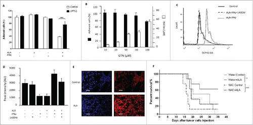
Figure 5. The toxic effect of ALA plus IFNγ on EMT-6H cells is due to peroxynitrite. (A) EMT-6H cells were treated with or without ALA (500 ng/mL) plus IFNγ (33 ng/mL) and 10 µM 1400 W or FeTPPS (5,10,15,20-tetrakis (4-sulfonatophenyl) porphyrinato iron (III) chloride; 150 µM), for 48 h. Peroxynitrite production was detected by adding luminol to cells and immediately measuring luminescence. Student's t test ****p < 0.0001. (B) Lung tissues were taken after the 3rd injection of ALA or saline solution as . The sections were stained with an anti-NO-tyrosine antibody (red). Scale bars: 20 μm. (C) EMT-6H cells were treated with or without ALA, IFNγ or FeTPPS for 48 h and cell viability was measured as described in Materials and Methods.
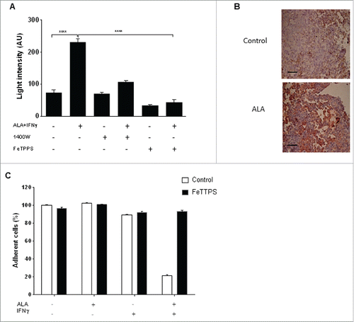
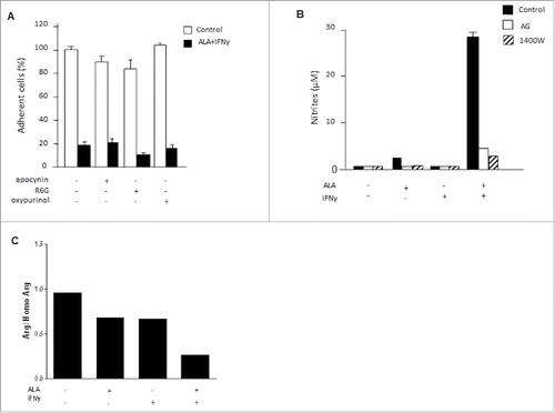
Figure 6. NOS II uncoupling was observed when EMT-6H cells were activated with ALA plus IFNγ. (A) EMT-6H cells were treated with or without NOX inhibitors: 300 µM apocynin, 0.3 µM rhodamine 6G, or 150 µM oxypurinol, for 1 h before adding ALA (500 ng/mL) plus IFNγ (33 ng/mL) for 48 h. Cell viability was detected as described in Materials and methods. Data from one representative of three independent experiments in triplicate are shown. (B) EMT-6H cells were treated for 1 h with 0.5 mM AG or 10 µM 1400 W, then for 48 h with or without ALA and IFNγ. Nitrite accumulation was measured by the Griess assay. (C) EMT-6H cells were treated with or without ALA or IFNγ. L-arginine was measured in cell extracts by HPLC using homo-L-arginine as the internal standard. Data from one experiment representative of two are shown.