
Figures & data
Figure 1. Examples of typical expression patterns of TLR3, TLR7 and TLR8. (A)–(C) representing the same sample with normal epithelium (NE), low-grade dysplasia (LGD), high grade dysplasia (HGD) and adenocarcinoma (CA) marked in (A). Gradual increase is found through normal epithelium–metaplasia–dysplasia sequence. (D)–(F) show intestinal type metaplasia (top), normal epithelium (middle) and adenocarcinoma (bottom). Intestinal metaplasia in TLR3, 7 show basal polarization, whereas TLR8 is expressed more diffusely. Expression pattern of all studied TLRs in adenocarcinoma is diffuse extending homogenously throughout the cell cytoplasm with no apparent basal polarization. (G)–(I) show intestinal metaplasia (left) and gastric type metaplasia (right). Gastric metaplasia presented a strong polarized staining to the basal cytoplasm in all studied TLRs. Magnifications 6× and 20× were used.
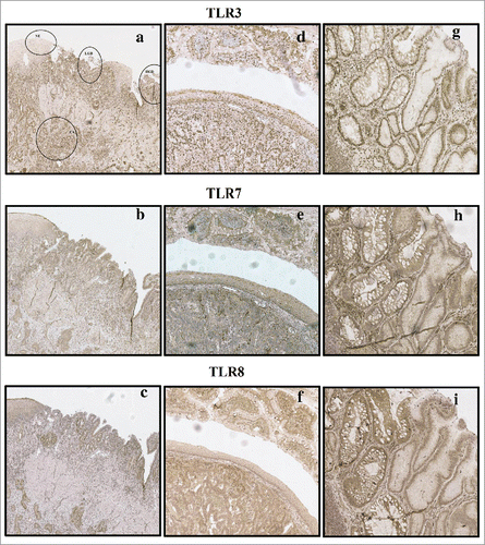
Figure 2. Positive TLR8 nuclear expression in immnohistochemistry (A), immunofluorescence photomicrograph obtained by confocal microscopy (B) and DNA-specific TO-PRO2 as a positive control (C). Negative TLR8 nuclear expression in immunohistochemistry (D) and immunofluorescence (E). Nuclei are visible with DNA-specific TO-PRO2 staining (F). Magnification of 40x was used.
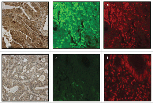
Table 1. Baseline characteristics of TLR3, 7 and 8 expression in normal esophageal squamous epithelium and in different esophageal lesions. Intensity was assessed with a 4-point scale from negative (0) to strong intensity (3). The extent of the staining was expressed as percentage of positive cells and positive cell nuclei (0–100%). Histoscore is counted by multiplying intensity with the percentage of positive cells (0–300) Values are presented as mean, median and interquartile range (IQR). Statistically significant differences are shown for histoscores and nuclear TLR expression. Letters are placed to indicate the lesion with higher TLR expression in each comparison.
Figure 3. Kaplan–Meier survival curve in esophageal adenocarcinoma stratified by nuclear expression of TLR8 in the cancer cells.
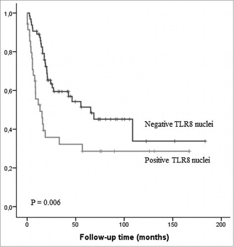
Table 2. The relationships between TLR3, 7 and 8 histoscores compared to clinicopathological variables in esophageal adenocarcinoma.
Table 3. Association of presence of nuclear TLR8 expression and clinicopathological variables in esophageal adenocarcinoma. Significant p values are shown in bold.