Figures & data
Figure 1. PD-L1 IHC-staining patterns: expression was noticed in the tumor cells (A), in tumor-associated immune cells (B) or in both cell types (C; white arrow head: PD-L1-positive tumor cells, black arrow heads: PD-L1-positive immune cells). The proportion of positive cells varied with some cases showing few positive cells (A1–C1) while others showed mostly positive cells (A2–C2) (IHC PD-L1, Ab 5H1, 560x).
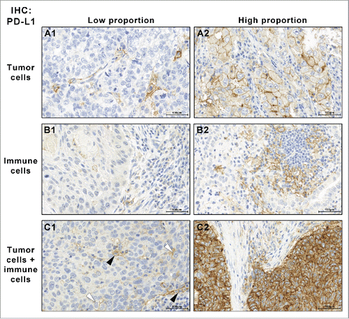
Figure 2. Tumor-associated PD-L1-positive immune cells stain positive for macrophage marker proteins. PD-L1-positive immune cells (black arrow heads) were encountered in combination with PD-L1-positive tumor cells (A1) or as the only PD-L1-positive cell population (B1). In all cases, PD-L1 was positive in immune cells that were also positive for macrophage marker proteins CD163 (A2, B2) and CD68 (Figs. S1, 2).
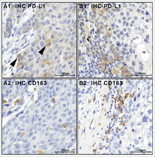
Figure 3. PD-L1 IHC positive (+) and negative (–) tumor cells (TC) and immune cells (IC) in pulmonary adenocarcinoma and squamous cells carcinoma. For each subgroup, absolute numbers and relative proportions are indicated. PD-L1-positive ICs are frequent and may occur isolated or in combination with positive TCs.
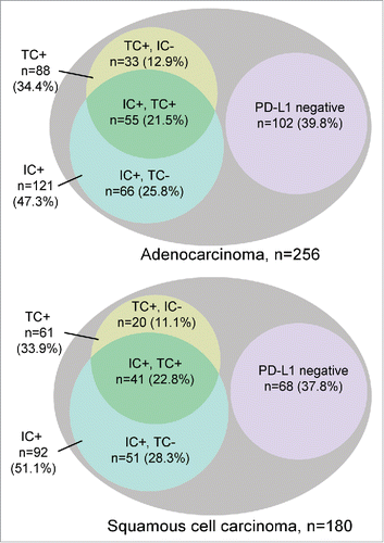
Table 1. Clinical data of the 436 NSCLC patients.
Table 2. Summary of PD-L1 protein expression, (A) in the tumor cells (TC) and (B) in tumor-associated immune cells (IC). Cases were classified PD-L1 IHC “positive” if ≥ 1% of the TCs or ICs were stained.
Table 3. In adenocarcinoma PD-L1 expression in TC was found in both early NSCLC (UICC stage I/II) and advanced NSCLC (UICC stage III/IV). Significant associations were found for KRAS, TP53 and STK11 mutations in univariate regression analysis (depicted) and in multivariate analysis (). All other investigated parameters did not show significant associations (Tables S1, 2).
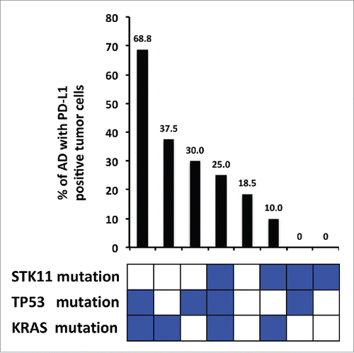
Figure 4. Percentage of PD-L1 expression in adenocarcinomas and mutational status of significantly associated genes. The combination of TP53 mutation, KRAS mutation and STK11 wildtype is associated with the highest percentage of PD-L1 expression in adenocarcinoma tumor cells. Conversely, STK11 mutations in the absence of TP53 and KRAS mutations are associated with the lowest percentage.