Figures & data
Figure 1. Lenalidomide therapy enhances the antigen-specific immune response of CD8+ T cells from patients with multiple myeloma. The number of patients with specific or no specific immune response treated with or without lenalidomide is shown.
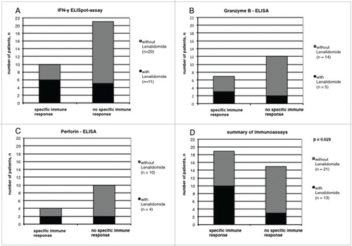
Figure 2. Impact of lenalidomide therapy on the expression of T-cell markers. Shown is the expression of (A) CD45RA, (B) CD28, (C) CCR7 and (D) CD279 (E) CD38, (F) CD154, (G) HLA-DR, (H) CD57 on CD8+ T cells (in % of all CD8+ T cells) from patients with MM treated with or without lenalidomide, analyzed by flow cytometry.
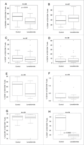
Figure 3. Impact of lenalidomide therapy on the CD4+CD127dimCD25high Foxp3+regulatory T-cell compartment. Shown is the percentage of CD4+CD127dimCD25highFoxp3+regulatory T cell of all CD4+ T cells in patients with MM treated with or without lenalidomide, analyzed by flow cytometry.
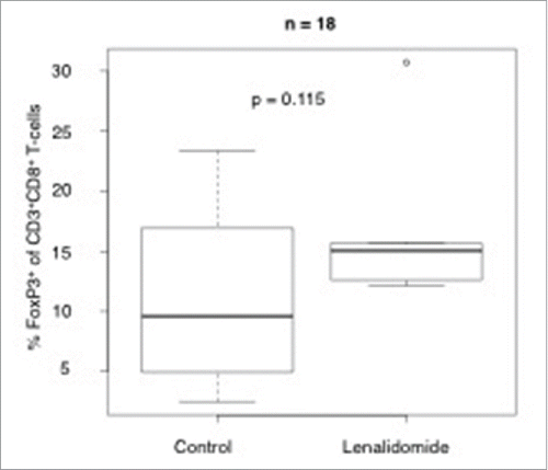
Figure 4. Representative EliSpot-analysis. The figure shows a representative EliSpot analysis from a lenalidomide refractory patient with T cells demonstrating an enhanced antigen-specific T-cell response against the myeloma antigen HM1.24 after in vitro incubation with lenalidomide.
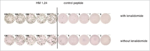
Table 1. Patient characteristics (newly diagnosed patients).
Table 2. Patient characteristics (patients with lenalidomide-refractory multiple myeloma).
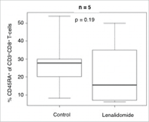
Figure 5. Impact of lenalidomide in vitro in patients with refractory multiple myeloma on the expression of T-cell markers. The plot shows the expression of CD45RA on CD8+ T-cells (in % of all CD8+ T cells) from patients with refractory MM analyzed by flow cytometry.