Figures & data
Table 1. Clinicopathological characteristics of patients and tumors (N = 758).
Figure 1. Metastasis-free survival according to PDL1 expression in soft-tissue sarcomas. (A) Kaplan–Meier MFS curves in all patients (N = 470). (B) Similar to (A), but in the learning set (N = 235) and according to PDL1 expression. (C) Similar to (B), but in the validation set (N = 235). (D) Similar to (B), but in the pooled learning and validation sets (N = 470).
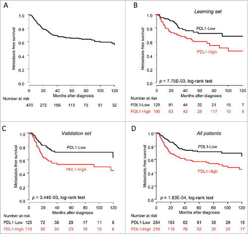
Table 2. Correlations of PDL1 expression with clinicopathological variables (N = 758).
Table 3. Univariate and multivariate prognostic analyses for metastasis-free survival (N = 470).
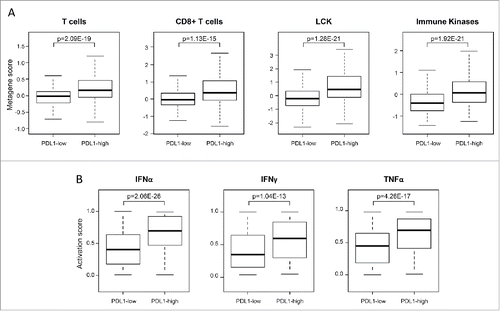
Figure 2. Correlations of PDL1 expression with immune features in soft-tissue sarcomas. (A) Metagene expression scores in all STS (N = 758) reported as a box plot according to PDL1 expression status for T cells and CD8+ T-cells metagenes and two prognostic immune kinase gene expression signature (LCK and immune kinases). (B) Similar to (A), but showing the probability of activation of immune pathways including IFNα, IFNγ, and TNFα pathways. The p-values (Student's t-test) are indicated.