Figures & data
Figure 1. IL4I1 is mainly expressed by myeloid cells and correlates with disease progression in Ret mice (A and D) IL4I1 activity in cervLN, spleen (A) or tumor fractions (D) from WT (white) or Ret (black) mice. (B) Pearson correlation of IL4I1 activity from spleen of Ret mice depending on the tumor stage. (C and E) IL4I1 expression was measured by qRT-PCR in purified CD11b+ or CD11b− fractions isolated from spleen (C) or primary tumors (E) of Ret mice. Experiments were performed from Ret mice at different stages of melanoma development (A and B), or from 3 to 6-mo old mice exhibiting distant metastasis, respectively (C–E). Data were pooled from at least three independent experiments. *p < 0.05; ****p < 0.0001.
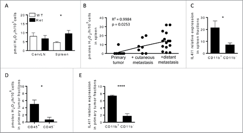
Figure 2. IL4I1 favors melanoma progression and metastasis formation with no direct effect on tumor cell proliferation and angiogenesis. (A–E) Melanoma progression in Ret (black line, n = 28) or RetIL4I1KO (gray line, n = 32) mice was evaluated once a week over a 3-mo period. Time courses of primary tumor (A), facial (B) or dorsal (C) metastasis onset. Frequency of 3-mo old mice with distant metastasis (D) or vitiligo (E) are shown. Mantel-Cox test (A–C). (F) Illustration and quantification of the CD34 vessel staining in primary tumors of 3-mo old Ret or RetIL4I1KO mice. Scale bar, 100 µm. (G) Growth comparison of B16-F10 cells seeded in E-plates wells with or without recombinant IL4I1. (H) Proportion of KI67+ cells among CD45− cells within the primary tumors of 3-mo old Ret and RetIL4I1KO mice. Data were pooled from at least three experiments. *p < 0.05.
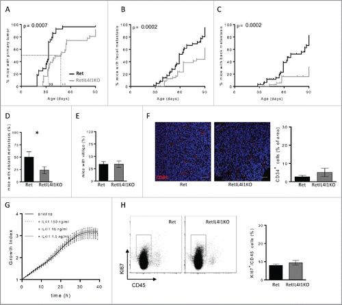
Figure 3. Remodeling of immune cell infiltration in primary tumor of mice lacking IL4I1. (A) Proportions and absolute numbers of CD45+ cells within primary tumors of 3-mo old Ret and RetIL4I1KO mice. (B) Distribution of immune cells infiltrating Ret (top) and RetIL4I1KO (low) primary tumors. (C) Proportion and absolute numbers of myeloid cells. (D–F) Absolute numbers of DC (CD11c+), MФ (CD11b+CD11c−Ly6C−/lowLy6G−), M-MDSC (CD11b+CD11c−Ly6ChighLy6G−) and PMN-MDSC (CD11b+CD11c−Ly6CintLy6G+) (D) and also CD19+ B cells and TCR+ cells (E) subdivided into CD4+ or CD8+ T cells (F). Representative gating strategies are shown (D, E, F; left). Data are pooled of at least three independent experiments. Mean and SEM. *p < 0.05; **p < 0.01; ***p < 0.001.
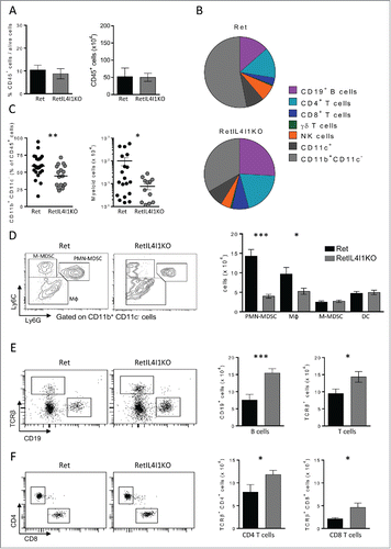
Table 1. Quantification of immune populations infiltrating the primary tumor (%).
Figure 4. IL4I1 deficiency improves anti-tumor properties of CD4+ and CD8+ T cells, but not the generation of tumor-specific antibodies. (A) Flow-cytometric analysis of the presence of antitumor antibodies in the sera of 3-mo old Ret and RetIL4I1KO mice. (Left) Representative histograms (light gray, staining with the secondary antibody alone; black, Ret mice serum; dark gray, RetIL4I1KO mice serum), (right) MFI ratios were calculated by dividing the MFI obtained with a given serum by the MFI obtained with the secondary antibody. (B-F) Frequency (%) of CD4+ or CD8+ T cells expressing KI67 (B and D) or IFNγ (Cand E) and the proportion of CD8+ CD107a+ within the primary tumor. Graphs depict the mean ± SEM from at least two independent experiments. **p < 0.01; ****p < 0.00001.
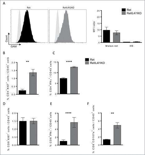
Table 2. Patients and melanoma characteristics.
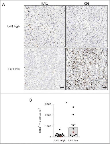
Figure 5. Primary tumors poorly infiltrated by IL4I1+ cells exhibit an enhanced density of CD8+ T cells in melanoma patients (A) Representative stainings of CD8+ and IL4I1+ cells infiltrating melanoma tumors. Scale bar, 100 µm. (B) Quantifications of CD8+ T cell density in low (n = 9) or high (n = 11) IL4I1-infiltrated tumors (corresponding to < or > of the median value of IL4I1+ cell density). Each point corresponds to a primary tumor. Red symbols display the representative images in A. Bars depict mean ± SEM. Unpaired t test; *p < 0.05.