
Figures & data
Figure 1. Injection of Breg ameliorates GVHD. Lethally irradiated BALB/c recipients (8 Gy) were transplanted with 5 × 10⁶ TCD-BM derived from B6 mice (n = 11) or with TCD-BM plus spleen T cells (n = 14). Breg (3 × 10⁶) was injected i.p. into T cell recipients at the time of transplantation (n = 15). (A) The survival of BMT recipients was monitored over time (p < 0.01). (B) Recipient mice were assessed every 2 d for clinical severity of GVHD; clinical scores are shown (p < 0.05). (C) Histopathology of skin, liver, intestine, and colon of BMT recipients 14 d after transplantation (original magnification × 200). Upper panel is TCD-BM + T cells + Breg group; middle panel is TCD-BM + T cells group; and lower panel is TCD-BM group. (D) Pathologic damage in the intestine, colon, skin, and liver was assessed using a semi-quantitative scoring system, as described in materials and methods. Results are representative of three independent experiments. Data are mean ± SEM.
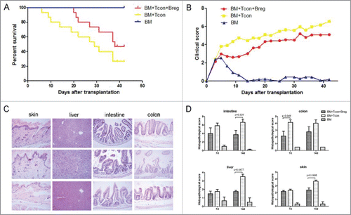
Figure 2. Bregs modulate Th cell balance in GVHD. Lethally irradiated BALB/c recipients were transplanted with TCD-BM derived from B6 mice or with TCD-BM plus spleen T cells. Breg (3 × 10⁶) was injected into T cell recipients at the time of transplantation. (A) Flow cytometry analysis of intracellular IL-4 on CD4+ T cells from spleen on indicated days. (B) TNF-α and IFNγ concentrations were determined in the serum of recipient mice 7 d after BMT. (C) Flow cytometry analysis of transcription factor T-bet, GATA3, and RORγt on CD4+ T cells from spleen (SP) of recipients with and without Breg injection on the indicated days. (D) Th2/Th1 and Th2/(Th1+Th17) ratios in peripheral blood, bone marrow, and spleen of recipients with and without Breg injection on the indicated days. Results are representative of three independent experiments with three mice per group per experiment. Data are mean ± SEM. *p < 0.05, **p < 0.01, ***p < 0.001.
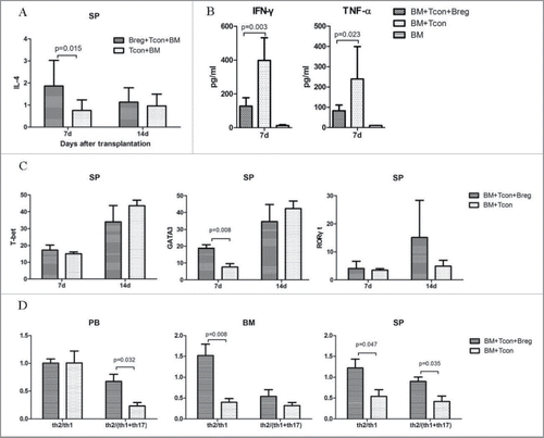
Figure 3. Bregs attenuate GVHD via Tregs in vivo. (A) Flow cytometry analysis results showing representative percentages of donor-derived peripheral blood and bone marrow T cells expressing Foxp3 on days 7 and 14. (B) Treg/Th1 and Treg/(Th1+Th17) ratios in peripheral blood of recipients with and without Breg injection on the indicated days. (C) Immunohistochemical staining to assess the number of T-bet-, GATA3-, RORγt-, and Foxp3-expressing cells in the skin, intestine, colon, liver, and lung on days 7 and 14. Results are representative of three independent experiments with three mice per group per experiment. Data are mean ± SEM. *p < 0.05, **p < 0.01. ns, p > 0.05.
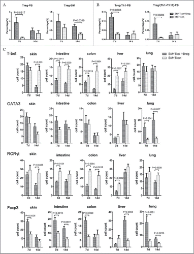
Figure 4. Bregs preserve the (T)cell-mediated GVL effect while inhibiting GVHD. Lethally irradiated BALB/c recipients were transplanted with B6 TCD-BM, with or without T cells, and challenged or not with leukemia cells (5 × 10⁵/mouse). Bregs were injected at the time of transplantation. (A) Survival was monitored over time. (B) Recipient mice were assessed every 2 d for clinical severity of GVHD; clinical scores are shown. (C) Mean percentage initial body weight of recipients over the first 30 d. (D) A representative dot plot for GFP+ leukemia cells. (E) GFP+ leukemia cells in peripheral blood and bone marrow were counted by flow cytometry on days 7 and 14. (F) On day 14, mice from different groups were killed, and autopsy was performed to observe the enlargement of liver (upper panel) and spleen (lower panel). (G) Comparable mouse general physical status was found in the Breg injection group and bone marrow-transplanted group. From left to right, TCD-BM + T cells + leukemia cells + Breg, TCD-BM + T cells + leukemia cells, TCD-BM + leukemia cells, TCD-BM. Results are representative of three independent experiments with three mice per group per experiment. Data are mean ± SEM. *p < 0.05, **p < 0.01, ***p < 0.001, ****p < 0.0001.
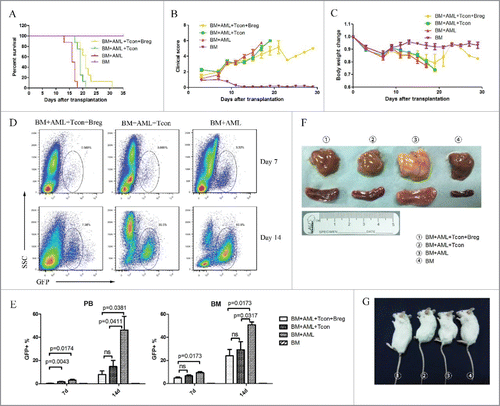
Table 1. Patient and donor characteristics.
Figure 5. Outcome of allogeneic stem cell transplantations in the cohort of patients. (A) Grades II–IV acute GVHD, (B) relapse, (C) non-relapse mortality, (D) disease-free survival, and (E) overall survival. Patients receiving a high dose of Bregs in allografts (more than or equal to 1.63 × 10⁷ Breg cells infused/kg) (n = 37): green line; subjects receiving a high dose of Bregs in allografts (less than 1.63 × 10⁷ Breg cells infused/kg) (n = 37): blue line.
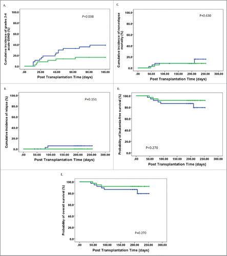
Table 2. Transplant outcome of patients that underwent allo-HSCT.