Figures & data
Figure 1. Generation and molecular characterization of patient-derived xenograft (PDX) models of non-small-cell lung cancer. (A) Hematoxylin and eosin (H&E) staining and immunohistochemistry detection of human leukocyte antigen (HLA), E-cadherin and vimentin in tumor sections from both the first and second passages of PDX mice for patient P2 (see from ). Although HLA+ cells from PDX mice also expressed E-cadherin, cells from both passages were negative for vimentin. (B) H&E staining and immunohistochemistry detection of HLA, E-cadherin and vimentin in sections from liver, spleen and subcutaneous (s.c.) tissue. All sections were from a single mouse among the third passage of PDX mice for patient P1 (see from ). Scale bar = 20 μm.
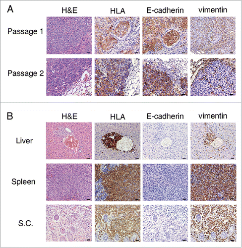
Table 1. Clinical information of the patients and characteristics of the corresponding PDX models.
Figure 2. Sections of tumors from eight patient-derived xenograft (PDX) models. Representative images correspond to tumors derived from eight patients; all sections were stained with hematoxylin and eosin (H&E) and antibodies against human leukocyte antigen (HLA), PSCA and MUC1. The passage numbers of each PDX for the patients were indicated. The negative controls (NC) are the liver tissues from a same mouse of third passage of PDX for patient P3. Scale bar = 20 μm. PSCA and MUC1 detection results are also shown in .
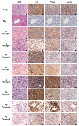
Figure 3. Construction of anti-prostate stem cell antigen (PSCA) and anti-mucin 1 (MUC1) chimeric antigen receptor (CAR) T cells. (A) Structures of the genes used for lentiviral transfection. GFP, control without CAR; CAR-PSCA, anti-PSCA CAR; CAR-MUC1, anti-MUC1 CAR. (B) Representative flow cytometric analysis of transfected T cells. (C) Reverse transcription-PCR detection of the following: β-ACTIN in wild type (WT), CAR-PSCA and CAR-MUC1 T cells (left); anti-PSCA scFv in WT and CAR-PSCA T cells and CAR-PSCA vector as a positive control (middle) and anti-MUC1 scFv in WT and CAR-MUC1 T cells and CAR-MUC1 vector as a positive control (right).
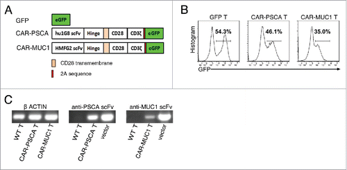
Figure 4. T cells expressing the prostate stem cell antigen (PSCA) or mucin 1 (MUC1) chimeric antigen receptor (CAR) specifically killed PSCA+ or MUC1+ lung cancer cell lines, respectively, in vitro. (A) Flow cytometric analysis of PSCA expression on A549, H23 and H460 cell lines. (B) Percentages of lung cancer line cells killed by GFP T cells and CAR-PSCA T cells at the indicated effector (E): target (T) ratios. The ratios were of the absolute number of CAR T cells vs target cells (corrected for transduction efficiency). T cells were co-cultured with A549GL, H460GL, H23GL or H23-PSCA-GL cells for 18 h, and luciferase activities were measured using a D-luciferin substrate. % of killing = % (total activities without T cells − activities with T cells)/ total activities without T cells. Data were representative of killing assays using T cells from three different donors. (C) Results of enzyme-linked immunosorbent assays (ELISAs) to detect IL-2 and IFNγ in the supernatants of co-cultures at a E:T ratio of 1:1. (D) Flow cytometric analysis of MUC1 expression on A549, H23 and H460 cell lines. (E) Percentages of lung cancer line cells killed by GFP T cells and CAR-MUC1 T cells at the indicated E:T ratios. (F) Results of ELISAs to detect IL-2 and IFNγ in the supernatants of cocultures at an E:T ratio of 1:1. Data were representative of killing assays using T cells from three different donors. (G) Post-transfection GFP-luciferase (GL) expression was detected in A549GL, H23GL and H460GL cell lines by flow cytometry. GFP served as a marker of luciferase expression. (H) Flow cytometric detection of PSCA (left) and MUC1 (right) in H23GL cells after lentiviral transduction. Error bars denote standard errors of the means, and groups were compared using the unpaired t-test. *p < 0.05, **p < 0.01, ***p < 0.001.
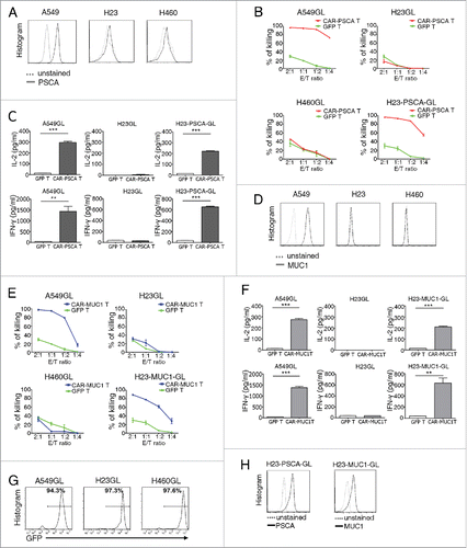
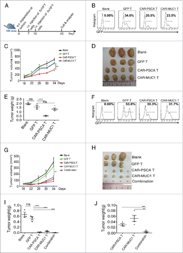
Figure 5. Prostate stem cell antigen chimeric antigen receptor (CAR-PSCA) expressing T cells inhibit the growth of non-small-cell lung cancer (NSCLC) and exhibit synergistic efficacy with mucin 1 CAR (CAR-MUC1) expressing T cells against NSCLC in patient-derived xenograft (PDX) models. (A) Diagram of the experiment with primary NSCLC tumors from patient P2 or P8 in NSI mice. Mice were inoculated subcutaneously with dissected tumor masses from patient P2 or P8 (2 mm × 2 mm), infused with 5 × 10⁶ total T cells on days 7 and 10, and culled on day 40 for tumor analysis. (B–E) Results from the PDX model of patient P2. (B) T cells were analyzed for transfection efficiency before infusion into PDX mice of patient P2. (C) Tumor growth curves in groups treated with no T (n = 3), GFP T (n = 3), CAR-PSCA T (n = 4) or CAR-MUC1 T (n = 4) cells. (D) Tumors from mice treated with no T, GFP T, CAR-PSCA T or CAR-MUC1 T cells on day 40 are shown. One mouse from both the no T and GFP T groups died when tumors were small, which was not shown. (E) Comparison of the weights of tumors described in D. (F, J) Results from the PDX model of patient P8. (F) T cells were analyzed for transfection efficiency before infusion into PDX mice of patient P8. (G) Tumor growth curves in groups treated with no T (n = 3), GFP T (n = 3), CAR-PSCA T (n = 4), CAR-MUC1 T (n = 4), and combinatorial CAR T cells (n = 4). (H) Tumors from different groups in (G) on day 40 were shown. Also, both the no T and GFP T groups had one mouse died when tumors were small. (I) Comparison of the weights of tumors in H. (J) Tumors from CAR-PSCA T, CAR-MUC1 T and combinatorial groups were singled out for comparison. Error bars denote standard errors of the means, and groups were compared using the unpaired t-test. *p < 0.05, **p < 0.01, ***p < 0.001.