
Figures & data
Figure 1. Correlation of sPD-1 and sPD-L1 levels in individual patients with advanced pancreatic cancer (n = 41).
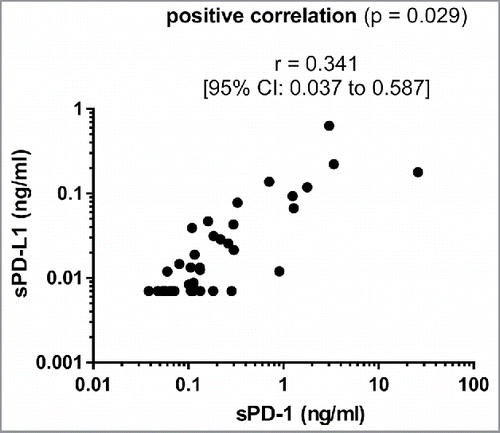
Table 1. Prognostic relevance of sPD-1 and sPD-L1.
Table 2A. Clinical characteristics of patients with high vs. low levels of sPD-1.
Table 2B. Clinical characteristics of patients with high vs. low levels of sPD-L1.
Figure 2. Overall survival for patients with high vs. low levels of sPD-1 or sPD-L1 (n = 41).
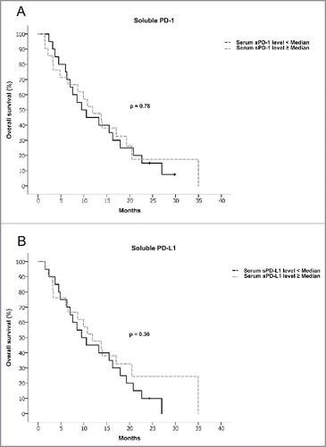
Figure 3. (A) Distribution of sPD-1 (left figure) and sPD-L1 (right figure) in patients with normal vs. elevated CRP. Differences in mean were tested using a Student's t-test, differences in distribution pattern were tested using an F-test. (B) Immunohistochemical staining of PD-L1 in controls (A, B) and exemplary pancreatic cancer tissue (C, D). Magnification = × 200. Scale bars indicate 50 µm. (C) Correlation analysis of sPD-L1 levels with tumoral CD3+ T cell infiltration (left figure) and tumoral PD-L1 expression (right figure) in advanced pancreatic cancer patients; Left figure: Correlation of sPD-L1 with tumoral CD3+ T cell infiltration (tCD3 count), differences between groups were tested using an F-test. Right figure: Correlation of sPD-L1 levels with tumoral PD-L1 expression, Pearson coefficient analysis was used to test for a potential correlation.
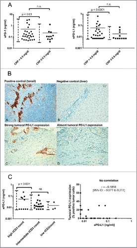
Table 3. Correlation of sPD-L1 levels with tumoral PD-L1 expression and tumoral CD3+ T cell infiltration in advanced pancreatic cancer.